

Ophthalmoplegic "migraine" or recurrent ophthalmoplegic cranial neuropathy: new cases and a systematic review
- PMID: 22241707
- PMCID: PMC3562350
- DOI: 10.1177/0883073811426502
Ophthalmoplegic "migraine" or recurrent ophthalmoplegic cranial neuropathy: new cases and a systematic review
Abstract
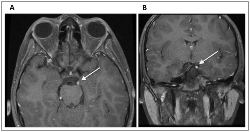
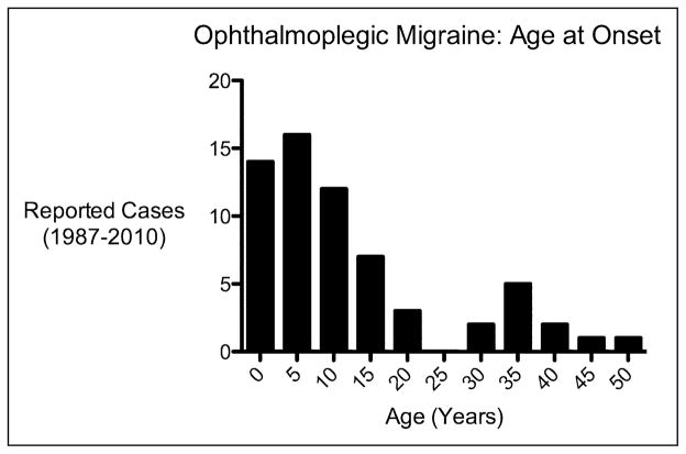
Ophthalmoplegic migraine is a poorly understood neurologic syndrome characterized by recurrent bouts of head pain and ophthalmoplegia. By reviewing cases presenting to our centers in whom the phenotype has been carefully dissected, and systematically reviewing all published cases of ophthalmoplegic migraine in the magnetic resonance imaging (MRI) era, this review sets out to clearly define the syndrome and discuss possible etiologies. We found that in up to one-third of patients, the headache was not migrainous or associated with migrainous symptoms. In three-quarters of the cases involving the third nerve, there was focal nerve thickening and contrast enhancement on MRI. Observational data suggest systemic corticosteroids may be beneficial acutely. The etiology remains unclear, but may involve recurrent bouts of demyelination of the oculomotor nerve. "Ophthalmoplegic migraine" is a misnomer in that it is probably not a variant of migraine but rather a recurrent cranial neuralgia. A more appropriate name might be "ophthalmoplegic cranial neuropathy."
Conflict of interest statement
The authors declared no potential conflicts of interest with respect to the research, authorship, and/or publication of this article.
Figures
Similar articles
-
Psychological therapies for the management of chronic and recurrent pain in children and adolescents.Cochrane Database Syst Rev. 2018 Sep 29;9(9):CD003968. doi: 10.1002/14651858.CD003968.pub5. Cochrane Database Syst Rev. 2018. PMID: 30270423 Free PMC article.
-
Antidepressants for pain management in adults with chronic pain: a network meta-analysis.Health Technol Assess. 2024 Oct;28(62):1-155. doi: 10.3310/MKRT2948. Health Technol Assess. 2024. PMID: 39367772 Free PMC article.
-
Diagnostic controversies in recurrent painful ophthalmoplegic neuropathy: single case report with a systematic review.Ital J Pediatr. 2022 Jun 3;48(1):82. doi: 10.1186/s13052-022-01274-x. Ital J Pediatr. 2022. PMID: 35659705 Free PMC article.
-
Non-pharmacological interventions for prophylaxis of vestibular migraine.Cochrane Database Syst Rev. 2023 Apr 12;4(4):CD015321. doi: 10.1002/14651858.CD015321.pub2. Cochrane Database Syst Rev. 2023. PMID: 37042522 Free PMC article.
-
What is the value of routinely testing full blood count, electrolytes and urea, and pulmonary function tests before elective surgery in patients with no apparent clinical indication and in subgroups of patients with common comorbidities: a systematic review of the clinical and cost-effective literature.Health Technol Assess. 2012 Dec;16(50):i-xvi, 1-159. doi: 10.3310/hta16500. Health Technol Assess. 2012. PMID: 23302507 Free PMC article.
Cited by
-
Approach to a patient with blepharoptosis.Neurol Sci. 2016 Oct;37(10):1589-96. doi: 10.1007/s10072-016-2633-7. Epub 2016 Jun 21. Neurol Sci. 2016. PMID: 27329276 Review.
-
[Differential diagnostics of chronic eye pain from a neurological perspective-What can also lie behind it].Ophthalmologie. 2023 Dec;120(12):1226-1232. doi: 10.1007/s00347-023-01958-7. Epub 2023 Nov 24. Ophthalmologie. 2023. PMID: 37999753 Review. German.
-
Recurrent alternating ophthalmoplegia with ipsilateral headache: unusual but possible manifestation of recurrent painful ophthalmoplegic neuropathy.Neurol Sci. 2020 Nov;41(11):3357-3360. doi: 10.1007/s10072-020-04502-6. Epub 2020 Jun 5. Neurol Sci. 2020. PMID: 32504277 No abstract available.
-
Headache for ophthalmologists: current advances in headache understanding and management.Eye (Lond). 2021 Jun;35(6):1574-1586. doi: 10.1038/s41433-021-01421-4. Epub 2021 Feb 12. Eye (Lond). 2021. PMID: 33580185 Free PMC article. Review.
-
Is ciliary muscle affected in migraine patients with aura and without aura?Med Sci Monit. 2015 Apr 28;21:1214-8. doi: 10.12659/MSM.893307. Med Sci Monit. 2015. PMID: 25919450 Free PMC article.
References
-
- McMillan HJ, Keene DL, Jacob P, Humphreys P. Ophthalmoplegic migraine: inflammatory neuropathy with secondary migraine? Can J Neurol Sci. 2007;34(3):349–355. - PubMed
-
- Bharucha DX, Campbell TB, Valencia I, Hardison HH, Kothare SV. MRI findings in pediatric ophthalmoplegic migraine: a case report and literature review. Pediatr Neurol. 2007;37(1):59–63. - PubMed
-
- Doran M, Larner AJ. MRI findings in ophthalmoplegic migraine: nosological implications. J Neurol. 2004;251(1):100–101. - PubMed
-
- Shin DJ, Kim JH, Kang SS. Ophthalmoplegic migraine with reversible thalamic ischemia shown by brain SPECT. Headache. 2002;42(2):132–135. - PubMed
-
- Headache Classification Subcommittee of the International Headache Society. The International Classification of Headache Disorders. Cephalalgia. (2) 2004;24(suppl 1):9–160. - PubMed
Publication types
MeSH terms
Substances
Grants and funding
LinkOut - more resources
Full Text Sources
