In situ formation of porous space maintainers in a composite tissue defect
- PMID: 22241726
- PMCID: PMC3288442
- DOI: 10.1002/jbm.a.34016
In situ formation of porous space maintainers in a composite tissue defect
Abstract
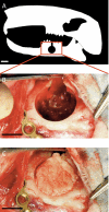
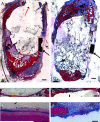
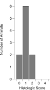
Reconstruction of composite defects involving bone and soft tissue presents a significant clinical challenge. In the craniofacial complex, reconstruction of the soft and hard tissues is critical for both functional and aesthetic outcomes. Constructs for space maintenance provide a template for soft tissue regeneration, priming the wound bed for a definitive repair of the bone tissue with greater success. However, materials used clinically for space maintenance are subject to poor soft tissue integration, which can result in wound dehiscence. Porous materials in space maintenance applications have been previously shown to support soft tissue integration and to allow for drug release from the implant to further prepare the wound bed for definitive repair. This study evaluated solid and low porosity (16.9% ± 4.1%) polymethylmethacrylate space maintainers fabricated intraoperatively and implanted in a composite rabbit mandibular defect model for 12 weeks. The data analyses showed no difference in the solid and porous groups both histologically, evaluating the inflammatory response at the interface and within the pores of the implants, and grossly, observing the healing of the soft tissue defect over the implant. These results demonstrate the potential of porous polymethylmethacrylate implants formed in situ for space maintenance in the craniofacial complex, which may have implications in the potential delivery of therapeutic drugs to prime the wound site for a definitive bone repair.
Copyright © 2012 Wiley Periodicals, Inc.
Figures


Similar articles
-
Evaluation of soft tissue coverage over porous polymethylmethacrylate space maintainers within nonhealing alveolar bone defects.Tissue Eng Part C Methods. 2010 Dec;16(6):1427-38. doi: 10.1089/ten.tec.2010.0046. Epub 2010 Jun 4. Tissue Eng Part C Methods. 2010. PMID: 20524844 Free PMC article.
-
Evaluation of antibiotic releasing porous polymethylmethacrylate space maintainers in an infected composite tissue defect model.Acta Biomater. 2013 Nov;9(11):8832-9. doi: 10.1016/j.actbio.2013.07.018. Epub 2013 Jul 25. Acta Biomater. 2013. PMID: 23891810
-
Use of porous space maintainers in staged mandibular reconstruction.Oral Maxillofac Surg Clin North Am. 2014 May;26(2):143-9. doi: 10.1016/j.coms.2014.01.002. Oral Maxillofac Surg Clin North Am. 2014. PMID: 24794263 Review.
-
Localized mandibular infection affects remote in vivo bioreactor bone generation.Biomaterials. 2020 Oct;256:120185. doi: 10.1016/j.biomaterials.2020.120185. Epub 2020 Jun 23. Biomaterials. 2020. PMID: 32599360 Free PMC article.
-
[Preparation and in vivo osteogenesis of acellular dermal matrix/dicalcium phosphate composite scaffold for bone repair].Zhongguo Xiu Fu Chong Jian Wai Ke Za Zhi. 2024 Jun 15;38(6):755-762. doi: 10.7507/1002-1892.202403059. Zhongguo Xiu Fu Chong Jian Wai Ke Za Zhi. 2024. PMID: 38918199 Free PMC article. Chinese.
Cited by
-
A composite critical-size rabbit mandibular defect for evaluation of craniofacial tissue regeneration.Nat Protoc. 2016 Oct;11(10):1989-2009. doi: 10.1038/nprot.2016.122. Epub 2016 Sep 22. Nat Protoc. 2016. PMID: 27658014
-
An experimental study on the impact of prosthesis temperature on the biomechanical properties of bone cement fixation.BMC Surg. 2023 Jul 5;23(1):191. doi: 10.1186/s12893-023-02079-3. BMC Surg. 2023. PMID: 37407954 Free PMC article.
-
Building bridges: leveraging interdisciplinary collaborations in the development of biomaterials to meet clinical needs.Adv Mater. 2012 Sep 18;24(36):4995-5013. doi: 10.1002/adma.201201762. Epub 2012 Jul 23. Adv Mater. 2012. PMID: 22821772 Free PMC article. Review.
-
Effects of Local Antibiotic Delivery from Porous Space Maintainers on Infection Clearance and Induction of an Osteogenic Membrane in an Infected Bone Defect.Tissue Eng Part A. 2017 Feb;23(3-4):91-100. doi: 10.1089/ten.TEA.2016.0389. Epub 2017 Jan 11. Tissue Eng Part A. 2017. PMID: 27998243 Free PMC article.
-
Advantages of Material Biofunctionalization Using Nucleic Acid Aptamers in Tissue Engineering and Regenerative Medicine.Mol Biotechnol. 2023 Dec;65(12):1935-1953. doi: 10.1007/s12033-023-00737-8. Epub 2023 Apr 5. Mol Biotechnol. 2023. PMID: 37017917 Review.
References
-
- Takushima A, Harii K, Asato H, Nakatsuka T, Kimata Y. Mandibular reconstruction using microvascular free flaps: a statistical analysis of 178 cases. Plast Reconstr Surg. 2001;108(6):1555–1563. - PubMed
-
- McLean JN, Moore CE, Yellin SA. Gunshot wounds to the face-acute management. Facial Plast Surg. 2005;21(3):191–198. - PubMed
-
- Vásconez HC, Shockley ME, Luce EA. High-energy gunshot wounds to the face. Ann Plas Surg. 1996;36(1):18–25. - PubMed
-
- Finch DR, Dibbell DG. Immediate reconstruction of gunshot injuries to the face. J Traum. 1979;19(12):965–968. - PubMed
-
- Becelli R, Renzi G, Perugini M, Iannetti G. Craniofacial traumas: immediate and delayed treatment. J Craniofac Surg. 2000;11(3):265–269. - PubMed
Publication types
MeSH terms
Grants and funding
LinkOut - more resources
Full Text Sources

