. 2012 Apr;40(7):e54.
doi: 10.1093/nar/gkr1263. Epub 2012 Jan 12.

Structural bias in T4 RNA ligase-mediated 3'-adapter ligation

Affiliations

Structural bias in T4 RNA ligase-mediated 3'-adapter ligation

Fanglei Zhuang et al. Nucleic Acids Res. 2012 Apr.

Abstract

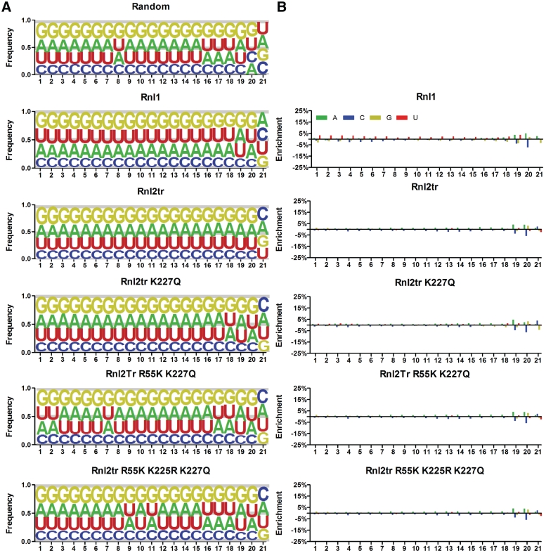
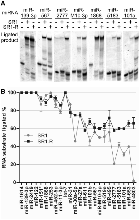
T4 RNA ligases are commonly used to attach adapters to RNAs, but large differences in ligation efficiency make detection and quantitation problematic. We developed a ligation selection strategy using random RNAs in combination with high-throughput sequencing to gain insight into the differences in efficiency of ligating pre-adenylated DNA adapters to RNA 3'-ends. After analyzing biases in RNA sequence, secondary structure and RNA-adapter cofold structure, we conclude that T4 RNA ligases do not show significant primary sequence preference in RNA substrates, but are biased against structural features within RNAs and adapters. Specifically, RNAs with less than three unstructured nucleotides at the 3'-end and RNAs that are predicted to cofold with an adapter in unfavorable structures are likely to be poorly ligated. The effect of RNA-adapter cofold structures on ligation is supported by experiments where the ligation efficiency of specific miRNAs was changed by designing adapters to alter cofold structure. In addition, we show that using adapters with randomized regions results in higher ligation efficiency and reduced ligation bias. We propose that using randomized adapters may improve RNA representation in experiments that include a 3'-adapter ligation step.


Figures

Figure 1.
Figure 1.
3′-adapter ligation efficiencies of miRNAs. (A) Each miRNA was incubated in a ligation reaction containing Rnl2tr with or without SR1 adapter. The ligation products were separated on 15% TBE–urea gels and visualized with SYBR Gold. Ligated products correspond to high molecular weight bands, which only appear in reactions with SR1 adapter. Unligated miRNAs and SR1 adapters remain as lower molecular weight bands. (B) The ligation efficiency of each miRNA was determined and plotted. The data are represented as the average ± standard deviation from two experimental replicates.
Figure 2.
Figure 2.
Scheme of in vitro ligation selection and sequencing library preparation. For each ligase selected library, an equal amount of 4 random RNA oligos containing a constant region (solid line), a randomized region (wavy line) and a known 3′-nt were combined to make a random oligo pool and used as substrates in a ligation reaction with pre-adenylated SR1 DNA adapter using a specific T4 RNA ligase. The ligated products were reverse transcribed and amplified to introduce the required primer regions for Ion Torrent sequencing. To determine the sequence content of the random RNA oligo pool, each of the four RNA oligos was sequenced independently. First, the oligos were poly A tailed for the random RNA oligo U, C and G or poly C tailed for the random RNA oligo A using poly(A) polymerase. The tailed RNA oligos were then reverse transcribed using primers complementary to the polymer tails (Supplementary Table S1). The cDNA libraries were amplified and processed in the same manner as the ligase selected libraries described above.
Figure 3.
Figure 3.
Nucleotide frequencies at each position in the randomized region of random and ligase selected libraries. (A) The nucleotide frequencies calculated from Ion Torrent sequencing runs of the random and ligase selected libraries were plotted in enoLOGOS format (40). The y-axis represents the frequency of each nucleotide proportional to the height of their representative letters, A, U, G and C. (B) The nucleotide frequencies of the ligase selected libraries were corrected to the frequencies in the randomized input library. The value of enrichment plotted on the y-axis is the normalized nucleotide frequency (RNnp) subtracting 0.25, ‘RNnp 0.25’. If (RNnp −0.25) of a nucleotide n at position p is equal to 0, it indicates the ligase doesn’t have preference for nucleotide n at position p. If RNnp −0.25) is greater or less than 0, it means that the nucleotide is preferred or not preferred at position p, respectively. The x-axis in A and B represents the position of nucleotides in the random region from 5′ to 3′.
Figure 4.
Figure 4.
Enrichment of RNA 3′-end predicted secondary structures in ligated libraries. Each sequence from the ligated libraries and random library was subjected to RNA CONTRAfold analysis. RNA structural predictions were classified based on the number of unpaired nucleotides at their 3′-end as labeled in the x-axis. A value of ‘42’ on the x-axis represents RNAs that lack any secondary structure according to CONTRAfold prediction. The value of enrichment was determined by the equation ‘(Observed–Expected)/Expected’, where ‘Observed’ is the percentage of a specific category in a ligated library and ‘Expected’ is the percentage of the same category in the random input library.
Figure 5.
Figure 5.
RNA-adapter cofold structures. Each sequence from random and ligated libraries was cofolded with the SR1 adapter using the Vienna RNAcofold. Based on the structural differences of predicted secondary structures at the ligation junction, 16 possible cofold structure classes are listed in (A). Each cofold structure class is numbered and presented in bracket and dot notion, in which brackets represent base pair(s) and dots represent unpaired nucleotide(s). The ‘&’ symbol represents the ligation junction between the RNA 3′-end and the adapter 5′-end. Multiple dots and brackets represent two or more unpaired or paired nucleotides in a row and the directionality of the brackets (open or closed) indicates the pairing orientation. Generalized schematic diagrams of corresponding cofolding structures are shown under the bracket and dot notation, in which RNA is in red and the DNA adapter is in black. The base pairings are shown as thin black lines. (B) Distribution of RNA and adapter cofold structures in simulated and sequenced libraries showing the percentage of library members assigned to each structural class. This distribution was used to calculate enrichment. (C) Enrichment of cofold structures in ligated libraries. The enrichment of each cofold structures was calculated using the equation, ‘(Observed–Expected)/Expected’, where ‘Observed’ is the percentage of a cofold structure in the ligated library and ‘Expected’ is the percentage of the corresponding structure in the random input library. Numbers on the x-axis correspond to the cofold structure classes in A and their schematic illustrations are shown under the numbers.
Figure 6.
Figure 6.
Comparison of miRNA ligation efficiencies using SR1 versus SR1-S adapter. (A) Sequences and predicted secondary structures of SR1 and SR1-S adapters. The underlined sequence is shared by both adapters. The secondary structures of SR1 and SR1-S are presented in bracket and dot form where brackets represent base paired nucleotides and dots represent unpaired nucleotides. (B) Ligation reactions of miRNAs with the SR1 or SR1-S adapter were performed using Rnl2tr. Ligation products were resolved in 15% TBE–urea gels and stained with SYBR Gold. (C) Ligation efficiency was calculated and plotted. The data points plotted represent average ligation efficiency ± standard deviation from two independent experiments.
Figure 7.
Figure 7.
Improving miRNA ligation efficiency using redesigned adapters. (A) Ligation of miRNAs with SR1 adapter or a new adapter specifically designed for each miRNA. Ligation reactions were performed using Rnl2tr. Ligation products were resolved in 15% TBE–urea gels, stained with SYBR Gold to visualize the nucleic acids. (B) Ligation efficiency was determined and plotted. The data points represent average ligation efficiency ± standard deviation from two independent experiments.
Figure 8.
Figure 8.
Improvement of miRNA ligation efficiencies using a randomized adapter, SR1-R. (A) Ligation reactions were performed with Rnl2tr and the SR1 or SR1-R adapter. Ligation products were resolved in 15% TBE–urea gels and stained with SYBR Gold to visualize the nucleic acids. (B) Ligation efficiencies of 24 miRNAs with the SR1 or SR1-R adapter were determined and plotted. The data are represented as the average ± standard deviation from two experimental replicates.
Figure 9.
Figure 9.
Improvement of miRNA ligation efficiency using a randomized adapter in the presence of mouse ES cell small RNAs. (A) Scheme of ligation reactions in the presence of mouse ES cell small RNAs. Each reaction contained 0.75 fmol of a 5′-32P labeled miRNA mixed with a 500-fold excess of mouse ES cell small RNAs and either the SR1 or the SR1-R adapter. Gray lines represent the ES cell small RNAs and the black line with an asterisk represents the radio-labeled miRNA. The SR1-R adapter is shown in black with a wavy line representing the random region at the 5′-end. Ligation products were resolved on 15% TBE–urea acrylamide gels, exposed to phosphor storage screens, and scanned. The ligated radio-labeled miRNA appears as a higher molecular weight band than unligated miRNA. (B) Representative results of ligation gels as described in A. (C) Comparison of ligation efficiency of miRNAs with the SR1 and SR1-R adapters. The intensity of ligated and unligated bands in each lane was quantified and ligation efficiencies were determined by calculating the percentage of ligated miRNA from the total miRNA. The data are represented as the average ± standard deviation ligation efficiency from two independent experimental replicates.

References

    1. Silber R, Malathi VG, Hurwitz J. Purification and properties of bacteriophage T4-induced RNA ligase. Proc. Natl Acad. Sci. USA. 1972;69:3009–3013. - PMC - PubMed
    1. Ho CK, Shuman S. Bacteriophage T4 RNA ligase 2 (gp24.1) exemplifies a family of RNA ligases found in all phylogenetic domains. Proc. Natl Acad. Sci. USA. 2002;99:12709–12714. - PMC - PubMed
    1. Cranston JW, Silber R, Malathi VG, Hurwitz J. Studies on ribonucleic acid ligase. Characterization of an adenosine triphosphate-inorganic pyrophosphate exchange reaction and demonstration of an enzyme-adenylate complex with T4 bacteriophage-induced enzyme. J. Biol. Chem. 1974;249:7447–7456. - PubMed
    1. Sugino A, Snoper TJ, Cozzarelli NR. Bacteriophage T4 RNA ligase. Reaction intermediates and interaction of substrates. J. Biol. Chem. 1977;252:1732–1738. - PubMed
    1. Amitsur M, Levitz R, Kaufmann G. Bacteriophage T4 anticodon nuclease, polynucleotide kinase and RNA ligase reprocess the host lysine tRNA. EMBO J. 1987;6:2499–2503. - PMC - PubMed