. 2012 Jan 15;26(2):126-36.
doi: 10.1101/gad.179895.111. Epub 2012 Jan 12.

Direct interaction of ligand-receptor pairs specifying stomatal patterning

Affiliations

Direct interaction of ligand-receptor pairs specifying stomatal patterning

Jin Suk Lee et al. Genes Dev. .

Abstract

Valves on the plant epidermis called stomata develop according to positional cues, which likely involve putative ligands (EPIDERMAL PATTERNING FACTORS [EPFs]) and putative receptors (ERECTA family receptor kinases and TOO MANY MOUTHS [TMM]) in Arabidopsis. Here we report the direct, robust, and saturable binding of bioactive EPF peptides to the ERECTA family. In contrast, TMM exhibits negligible binding to EPF1 but binding to EPF2. The ERECTA family forms receptor homomers in vivo. On the other hand, TMM associates with the ERECTA family but not with itself. While ERECTA family receptor kinases exhibit complex redundancy, blocking ERECTA and ERECTA-LIKE1 (ERL1) signaling confers specific insensitivity to EPF2 and EPF1, respectively. Our results place the ERECTA family as the primary receptors for EPFs with TMM as a signal modulator and establish EPF2-ERECTA and EPF1-ERL1 as ligand-receptor pairs specifying two steps of stomatal development: initiation and spacing divisions.


Figures
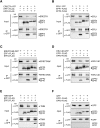
Figure 1.
Figure 1.
EPF1 and EPF2 associate with ERECTA and ERL1 but exhibit differential association with TMM in planta. Co-IP assays of epitope-tagged ligand–receptor pairs expressed in N. benthamiana leaves. (A) ERECTA (ERECTA-YFP) associates with EPF1 and EPF2. We observed that coexpression with EPFs results in reduced ERECTA-YFP signals, consistent with the previous report that full-length ERECTA protein is unstable (Shpak et al. 2005). (B) ERL1 (ERL1-YFP) associates with EPF1 and EPF2. (C) Kinase-deleted ERECTA (ERECTAΔK-GFP) associates with EPF1 and EPF2. (D) Kinase-deleted ERL1 (ERL1ΔK-GFP) associates with EPF1 and EPF2. (E) TMM does not associate with EPF1 but shows association with EPF2. (F) Control GFP does not associate with EPF1 or EPF2. The faint, higher molecular bands in the input blots of EPF1-Flag and EPF2-Flag likely represent variable unprocessed/intermediate precursor peptides. All experiments were repeated at least four times.
Figure 2.
Figure 2.
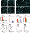
Production of bioactive, recombinant MEPF1 and MEPF2 peptides and detection of direct ligand–receptor binding. (A–H) Confocal images of wild-type cotyledon epidermis grown 5 d in a buffer solution (A,E; mock), 2.5 μM MEPF1 (B), or 2.5 μM MEPF2 (F), and transgenic seedlings expressing estrogen-inducible EPF1 (Est::EPF1) (C,D) or EPF2 (Est::EPF2) (G,H) grown in the absence (C,G; mock) or presence (D,H) of 5 μM estradiol. (Asterisks) Arrested stomatal precursors. Images were taken under the same magnification. Bar, 20 μm. (I–P) QCM and SPR analysis. (I,K) Average frequency shifts for representative QCM analysis using biosensor chips immobilized with ERECTAΔK-GFP (red), ERL1ΔK-GFP (green), TMM-GFP (blue), and GFP alone (gray) recorded sequentially after injection of MEPF1 (I) or MEPF2 (K) at serial dilution concentrations of active peptides. Error bars, SD. (J,L) Average SPR shifts with error bars (SEM). (M–P) Observed SPR kinetic sensograms fitted by least-square regression with a Langmuir adsorption model from representative SPR-binding assay upon injection (t = 0) of 300 nM refolded peptide solution of MEPF1 (M), MEPF2 (N), LURE2 (O), or buffer only (P) into biosensor chips immobilized with receptors as described above. SPR and QCM experiments were performed at least three times for each receptor using independent biosensor chips prepared for each method.
Figure 3.
Figure 3.
Epitope-tagged receptors are functional. Complementation of respective Arabidopsis mutant phenotypes by a series of epitope-tagged putative receptors. (A,B) Six-week-old plants of erecta erl1 erl2 triple loss-of-function mutant (A) and erecta erl1 erl2 expressing proERL1::ERL1-Flag (B). (C–E) Confocal microscopy of 12-d-old abaxial cotyledon epidermis of erecta erl1 erl2 (C), erecta erl2 (D), and erecta erl1 erl2 expressing proERL1::ERL1-Flag (E). The ERL1-Flag construct fully rescues both the growth defects (A,B) as well as the stomatal clustering (C,E) of erecta erl1 erl2, conferring an epidermal phenotype highly resembling that of erecta erl2. Note that the erl1 single mutant does not confer dwarfism or stomatal clustering. Brackets in D and E indicate arrested stomatal precursors typical of the erecta mutant background with functional ERL1 or ERL2. (F–H) Twelve-day-old abaxial cotyledon epidermis of tmm (F), proTMM::TMM-HA in tmm (G), and proTMM::TMM-YFP in tmm (H). Both proTMM::TMM-HA (G) and proTMM::TMM-YFP (H) rescue stomatal patterning defects seen in tmm (F). (I–M) Six-week-old plants of erecta (I) and erecta expressing proERECTA::ERECTA-YFP (J). (K–M) Twelve-day-old abaxial cotyledon epidermis of erecta (K) proERECTA::ERECTA-YFP in erecta (L), and proERECTA::ERECTA-Flag in erecta (M). The epitope-tagged receptors fully rescue the growth defects (I,J) and extra asymmetric division phenotype seen in erecta (K; brackets). Confirmed null (T-DNA knockout) alleles that do not accumulate any transcripts of respective receptor genes were used for all experiments. Full rescue of the erecta growth phenotype by proERECTA::ERECTA-Flag was described previously (Uchida et al. 2011). All confocal microscopy images were taken under the same magnification. Bars: whole-plant images, 5 cm; confocal microscopy, 20 μm.
Figure 4.
Figure 4.
Molecular interactions of ERECTA family RKs and TMM in vivo. Arabidopsis wild-type (wt) or null mutant seedlings expressing epitope-tagged receptors are indicated in each panel. In each panel, both inputs and co-IP fractions (IP α-GFP) were subjected to Western blot (WB) with anti-GFP (α-GFP) or anti-Flag (α-Flag) antibodies. (A) ERECTA associates with itself. (B) ERL1 associates with itself. (C) TMM does not associate with itself in vivo. Reciprocal co-IP assays resulted in no TMM bands in the co-IP fractions. (D) ERECTA associates with TMM. (E) ERL1 also associates with TMM. (F) ERECTA associates with ERL1, suggesting heteromerization among ERECTA family RKs. Immunoblots of inputs of TMM-HA and TMM-YFP yielded nonspecific bands. Co-IP experiments were performed at least three times.
Figure 5.
Figure 5.
Blocking ERECTA and ERL1 signaling with dominant-negative receptors confers specific insensitivity to EPF2 and EPF1, respectively. Confocal microscopy of liquid cultured 6-d-old abaxial cotyledons from wild type (WT; A,F,K), ERECTAΔK in erecta (B,G,L), epf2 (C,H,M), ERL1ΔK in erl1 (D,I,N), and epf1 (E,J,O). These seedlings were incubated with buffer only (mock; A–E), buffer with 2.5 μM MEPF1 (+MEPF1; F–J), or buffer with 2.5 μM MEPF2 (+MEPF2; K–O). (B,C) Both ERECTAΔK in erecta and epf2 mutation confer excessive entry asymmetric divisions (brackets). (D,E) Both ERL1ΔK in erl1 and epf1 mutation confer stomatal pairing (plus). (I) MEPF1 application confers an epidermis devoid of stomata but with arrested meristemoids (asterisks) in all genotypes except for ERL1ΔK in erl1, which produces paired stomata (plus). (L) MEPF2 application leads to severe inhibition of asymmetric division in all genotypes except for ERECTAΔK in erecta. Images were taken under the same magnification. Bar, 20 μm. Peptide application experiments were performed at least three times. For additional genotypes and quantitative analysis, see Supplemental Fig. 11.

References

    1. Abrash EB, Bergmann DC 2010. Regional specification of stomatal production by the putative ligand CHALLAH. Development 137: 447–455 - PubMed
    1. Abrash EB, Davies KA, Bergmann DC 2011. Generation of signaling specificity in Arabidopsis by spatially restricted buffering of ligand-receptor interactions. Plant Cell. 23: 2864–2879 - PMC - PubMed
    1. Baltus RE, Carmon KS, Luck LA 2007. Quartz crystal microbalance (QCM) with immobilized protein receptors: Comparison of response to ligand binding for direct protein immobilization and protein attachment via disulfide linker. Langmuir 23: 3880–3885 - PubMed
    1. Belkhadir Y, Chory J 2006. Brassinosteroid signaling: A paradigm for steroid hormone signaling from the cell surface. Science 314: 1410–1411 - PubMed
    1. Clark SE, Williams RW, Meyerowitz EM 1997. The CLAVATA1 gene encodes a putative receptor kinase that controls shoot and floral meristem size in Arabidopsis. Cell 89: 575–585 - PubMed

Publication types

MeSH terms