

Noninvasive imaging reveals inhibition of ovarian cancer by targeting CXCL12-CXCR4
- PMID: 22241961
- PMCID: PMC3257190
- DOI: 10.1593/neo.111076
Noninvasive imaging reveals inhibition of ovarian cancer by targeting CXCL12-CXCR4
Abstract
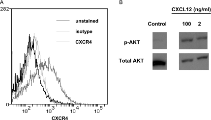
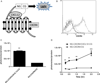
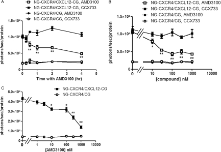
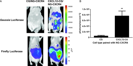
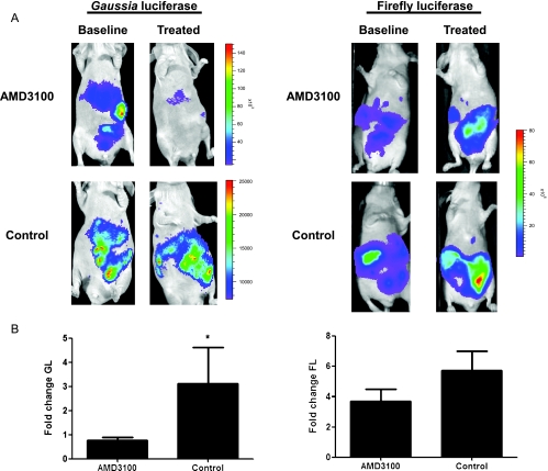
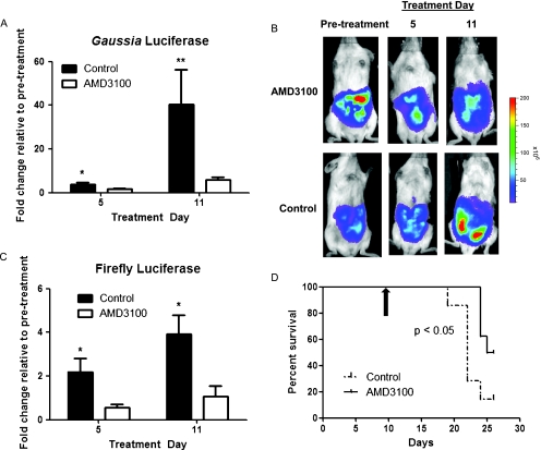
Patients with metastatic ovarian cancer continue to have a dismal prognosis, emphasizing the need for new strategies to identify and develop new molecular targets for therapy. Chemokine CXCL12 and its receptor CXCR4 are upregulated in metastatic ovarian cancer cells and the intraperitoneal tumor microenvironment. CXCL12-CXCR4 signaling promotes multiple steps in proliferation and dissemination of ovarian cancer cells, suggesting that targeted inhibition of this pathway will limit tumor progression. To investigate CXCL12-CXCR4 signaling in ovarian cancer and establish effects of inhibiting this pathway on tumor progression and survival, we designed a Gaussia luciferase complementation imaging reporter system to detect CXCL12 binding to CXCR4 in ovarian cancer cells. In cell-based assays, we established that the complementation imaging reporter could detect CXCL12 binding to CXCR4 and quantify specific inhibition of ligand-receptor interaction. We monitored CXCL12-CXCR4 binding and inhibition in a mouse xenograft model of metastatic human ovarian cancer by imaging Gaussia luciferase complementation and assessed tumor progression with firefly luciferase. Bioluminescence imaging studies in living mice showed that treatment with AMD3100, a clinically approved inhibitor of CXCL12-CXCR4, blocked ligand-receptor binding and reduced growth of ovarian cancer cells. Treatment with AMD3100 also modestly improved overall survival of mice with metastatic ovarian cancer. The Gaussia luciferase complementation imaging reporter system will facilitate further preclinical development and optimization of CXCL12-CXCR4 targeted compounds for treatment of ovarian cancer. Our research supports clinical translation of existing CXCR4 inhibitors for molecular therapy for ovarian cancer.
Figures
Similar articles
-
Imaging CXCL12-CXCR4 signaling in ovarian cancer therapy.PLoS One. 2013;8(1):e51500. doi: 10.1371/journal.pone.0051500. Epub 2013 Jan 23. PLoS One. 2013. PMID: 23372646 Free PMC article.
-
Role of CXCL12-CXCR4 axis in ovarian cancer metastasis and CXCL12-CXCR4 blockade with AMD3100 suppresses tumor cell migration and invasion in vitro.J Cell Physiol. 2019 Apr;234(4):3897-3909. doi: 10.1002/jcp.27163. Epub 2018 Sep 7. J Cell Physiol. 2019. Retraction in: J Cell Physiol. 2022 Mar;237(3):2005. doi: 10.1002/jcp.30517. PMID: 30191987 Retracted.
-
CXCL12/CXCR4 Axis-Targeted Dual-Functional Nano-Drug Delivery System Against Ovarian Cancer.Int J Nanomedicine. 2020 Aug 7;15:5701-5718. doi: 10.2147/IJN.S257527. eCollection 2020. Int J Nanomedicine. 2020. PMID: 32848392 Free PMC article.
-
The CXCL12 (SDF-1)/CXCR4 chemokine axis: Oncogenic properties, molecular targeting, and synthetic and natural product CXCR4 inhibitors for cancer therapy.Chin J Nat Med. 2018 Nov;16(11):801-810. doi: 10.1016/S1875-5364(18)30122-5. Chin J Nat Med. 2018. PMID: 30502762 Review.
-
Inhibition of CXCL12/CXCR4 axis as a potential targeted therapy of advanced gastric carcinoma.Cancer Med. 2017 Jun;6(6):1424-1436. doi: 10.1002/cam4.1085. Epub 2017 May 23. Cancer Med. 2017. PMID: 28544785 Free PMC article. Review.
Cited by
-
The interconnectedness of cancer cell signaling.Neoplasia. 2011 Dec;13(12):1183-93. doi: 10.1593/neo.111746. Neoplasia. 2011. PMID: 22241964 Free PMC article.
-
Plasmacytoid dendritic cells and regulatory T cells in the tumor microenvironment: A dangerous liaison.Oncoimmunology. 2013 May 1;2(5):e23887. doi: 10.4161/onci.23887. Oncoimmunology. 2013. PMID: 23762788 Free PMC article.
-
Quantification of ligand binding to G-protein coupled receptors on cell membranes by ellipsometry.PLoS One. 2012;7(9):e46221. doi: 10.1371/journal.pone.0046221. Epub 2012 Sep 26. PLoS One. 2012. PMID: 23049983 Free PMC article.
-
Modeling selective elimination of quiescent cancer cells from bone marrow.Neoplasia. 2015 Aug;17(8):625-33. doi: 10.1016/j.neo.2015.08.001. Neoplasia. 2015. PMID: 26408255 Free PMC article.
-
Tracking Tumor Colonization in Xenograft Mouse Models Using Accelerator Mass Spectrometry.Sci Rep. 2018 Oct 9;8(1):15013. doi: 10.1038/s41598-018-33368-0. Sci Rep. 2018. PMID: 30302019 Free PMC article.
References
-
- Kim S, Kim J, Kim S, Brantley E, Yun S, He J, Maya M, Zhang F, Wu Q, Lehembre F, et al. Macitentan (ACT-064992), a tissue-targeting endothelin receptor antagonist, enhances therapeutic efficacy of paclitaxel by modulating survival pathways in orthotopic models of metastatic human ovarian cancer. Neoplasia. 2011;13:167–179. - PMC - PubMed
Publication types
MeSH terms
Substances
Grants and funding
LinkOut - more resources
Full Text Sources
Medical