. 2012 Jan 10;8(1):17-23.
doi: 10.1021/ct200615k. Epub 2011 Nov 21.

Protecting High Energy Barriers: A New Equation to Regulate Boost Energy in Accelerated Molecular Dynamics Simulations

Protecting High Energy Barriers: A New Equation to Regulate Boost Energy in Accelerated Molecular Dynamics Simulations

William Sinko et al. J Chem Theory Comput. .

Abstract

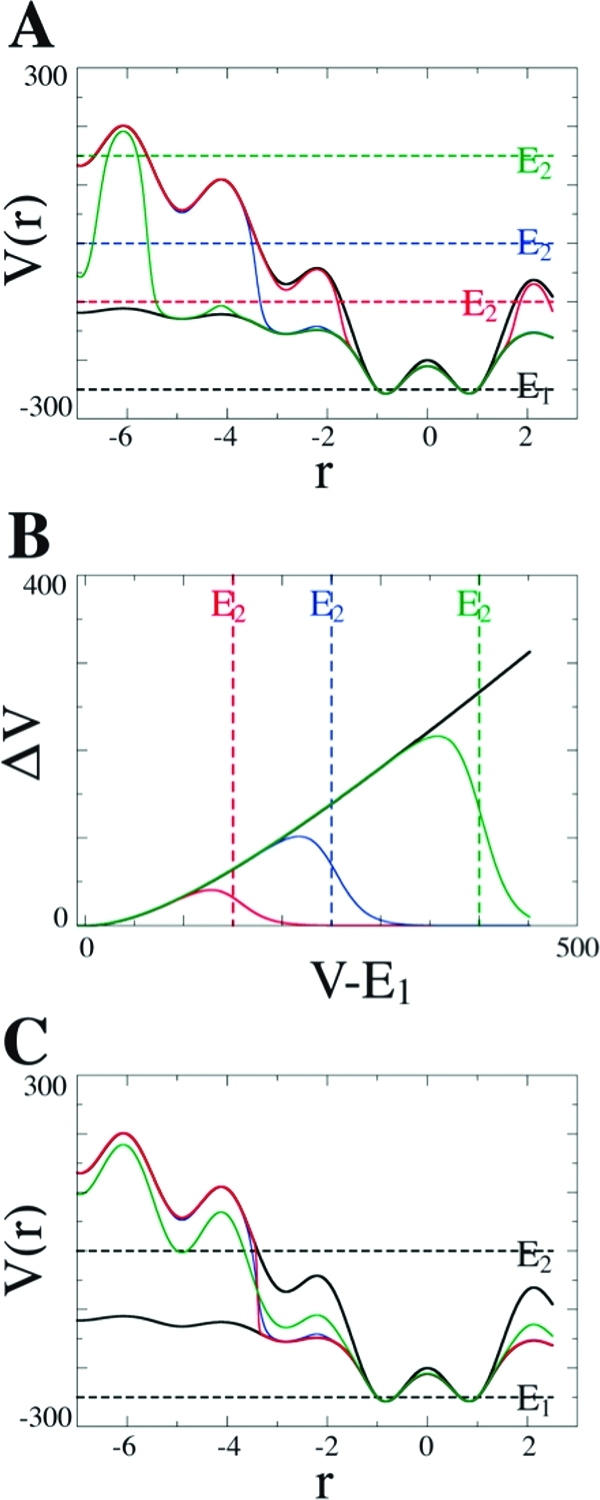
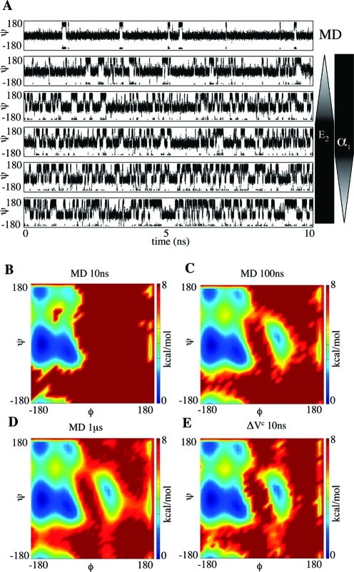
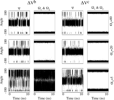
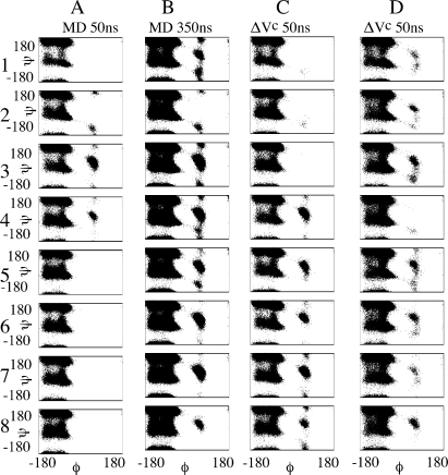
Molecular dynamics (MD) is one of the most common tools in computational chemistry. Recently, our group has employed accelerated molecular dynamics (aMD) to improve the conformational sampling over conventional molecular dynamics techniques. In the original aMD implementation, sampling is greatly improved by raising energy wells below a predefined energy level. Recently, our group presented an alternative aMD implementation where simulations are accelerated by lowering energy barriers of the potential energy surface. When coupled with thermodynamic integration simulations, this implementation showed very promising results. However, when applied to large systems, such as proteins, the simulation tends to be biased to high energy regions of the potential landscape. The reason for this behavior lies in the boost equation used since the highest energy barriers are dramatically more affected than the lower ones. To address this issue, in this work, we present a new boost equation that prevents oversampling of unfavorable high energy conformational states. The new boost potential provides not only better recovery of statistics throughout the simulation but also enhanced sampling of statistically relevant regions in explicit solvent MD simulations.


Figures

Figure 1
Figure 1
Hypothetical one-dimensional potential representing the effect of ΔVc. In all charts, α1 = 200 and E1 = −250. The upper and lower solid black lines represent the original potential and the modified potential generated with ΔVb, respectively. This color scheme is used throughout. (A) Effects of different parameters E2 (dashed colored lines) on the modified potential generated with ΔVc (solid colored lines). (B) Boost levels ΔVb (solid black line) and ΔVc (colored lines) as V(r) moves away from E1. For both A and B, α2=15 and E2 = −100 (red), 0 (blue), and 150 (green). (C) Effect of varying α2 parameter on ΔVc: α2= 3 (red), 15 (blue), and 75 (green) with E2 = 0.
Figure 2
Figure 2
Alanine dipeptide simulation results. (A) Ψ angle values obtained from cMD and five different aMD simulations. From top to bottom, aMD parameters were set to E2 = E1 + 15 and α1 = 5, E2 = E1 + 20 and α1 = 5, E2 = E1 + 25 and α1 = 5, E2 = E1 + 25 and α1 = 2.5, and E2 = E1 + 25 and α1 = 1.25. In all simulations, E1 and α2 were set to 10 and 5, respectively. Weighted free energy density plots obtained from cMD (B, C, D) and aMD with ΔVc (E). All values are in kcal/mol.
Figure 3
Figure 3
ψ and Ω angle values obtained from aMD simulations with boost potentials ΔVb and ΔVc. In all simulations, E1 = 10.0 and α1 were set as shown on the far right. Additional parameters for aMD with ΔVc were set to E2 = E1 + 15 and α2 = 5. All values are in kcal/mol.
Figure 4
Figure 4
Decalanine Φ–Ψ angles distribution obtained from cMD and aMD simulations. For the aMD simulations with ΔVc parameters were set to E1 = 74, E2 = E1 + 25, α2 = 5, α1 = 30 (C), and α1 = 15 (D). All values are in kcal/mol.
Figure 5
Figure 5
Principle component analysis obtained from decalanine MD simulations. (A) 50 ns of aMD simulation with ΔVc. Parameters were set to E1 = 74, E2 = E1 + 25, α2 = 5, and α1 = 15, same as in Figure 4D. (B) 50 ns of cMD simulation and (C) 350 ns of cMD simulation. Structures 1, 2, and 3 shown in yellow represent relevant populated states in PC subspace sampled by aMD and cMD.
Figure 6
Figure 6
(A) Relative free energy of binding between Ac2-L-Lys-D-Ala–-D-Ala and Ac2-L-Lys-D-Ala-D-Lac to vancomycin calculated from cMD (solid black line) and aMD with ΔVc (solid red line). A dashed line displays the experimental value, 4.1 kcal/mol. (B) Cumulative free energy curves calculated from simulations of 600 ps (left) and 1000 ps (right) per λ point. The ∗ shows points where there is no overlapping between error bars.

References

    1. Karplus M.; McCammon J. A. Nat. Struct. Biol. 2002, 9, 646. - PubMed
    1. Adcock S. A.; McCammon J. A. Chem. Rev. 2006, 106, 1589. - PMC - PubMed
    1. Hamelberg D.; Mongan J.; McCammon J. A. J. Chem. Phys. 2004, 120, 11919. - PubMed
    1. Voter A. F. Phys. Rev. Lett. 1997, 78, 3908.
    1. Markwick P. R.; Cervantes C. F.; Abel B. L.; Komives E. A.; Blackledge M.; McCammon J. A. J. Am. Chem. Soc. 2010, 132, 1220. - PMC - PubMed

LinkOut - more resources