. 2011 Dec;7(12):e1002452.
doi: 10.1371/journal.ppat.1002452. Epub 2011 Dec 29.

Inflammasome-dependent pyroptosis and IL-18 protect against Burkholderia pseudomallei lung infection while IL-1β is deleterious

Affiliations

Inflammasome-dependent pyroptosis and IL-18 protect against Burkholderia pseudomallei lung infection while IL-1β is deleterious

Ivonne Ceballos-Olvera et al. PLoS Pathog. 2011 Dec.

Abstract

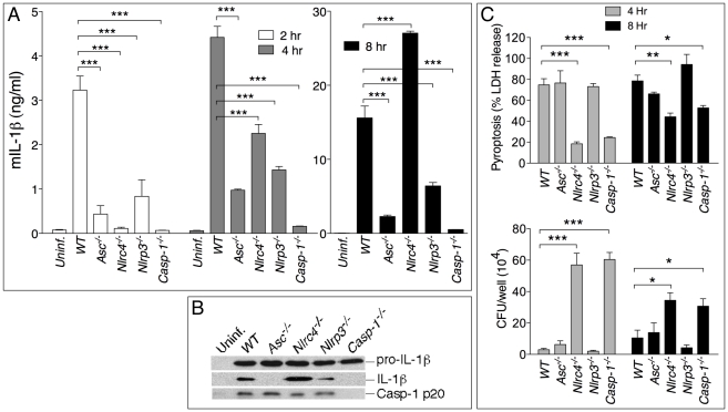
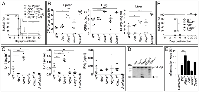
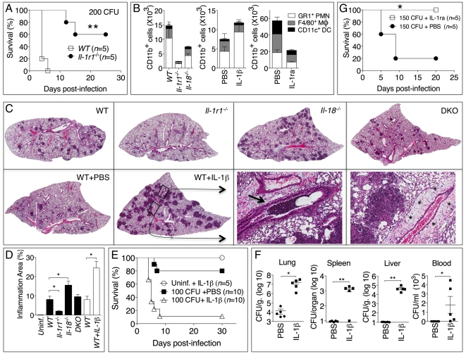
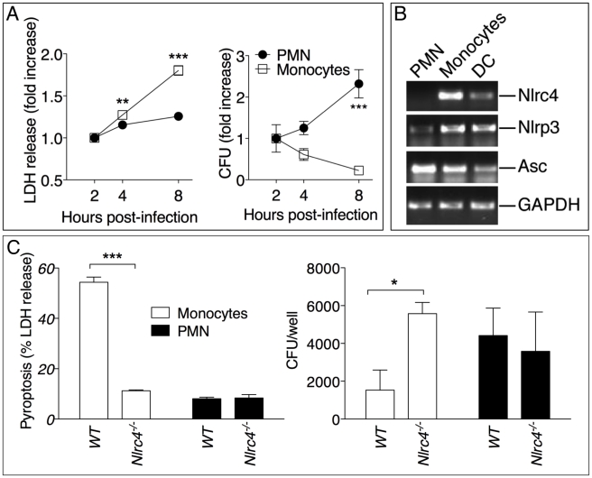
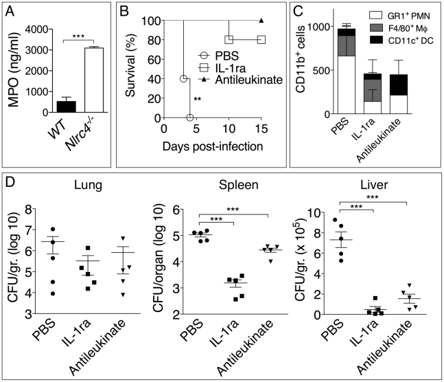
Burkholderia pseudomallei is a Gram-negative bacterium that infects macrophages and other cell types and causes melioidosis. The interaction of B. pseudomallei with the inflammasome and the role of pyroptosis, IL-1β, and IL-18 during melioidosis have not been investigated in detail. Here we show that the Nod-like receptors (NLR) NLRP3 and NLRC4 differentially regulate pyroptosis and production of IL-1β and IL-18 and are critical for inflammasome-mediated resistance to melioidosis. In vitro production of IL-1β by macrophages or dendritic cells infected with B. pseudomallei was dependent on NLRC4 and NLRP3 while pyroptosis required only NLRC4. Mice deficient in the inflammasome components ASC, caspase-1, NLRC4, and NLRP3, were dramatically more susceptible to lung infection with B. pseudomallei than WT mice. The heightened susceptibility of Nlrp3⁻/⁻ mice was due to decreased production of IL-18 and IL-1β. In contrast, Nlrc4⁻/⁻ mice produced IL-1β and IL-18 in higher amount than WT mice and their high susceptibility was due to decreased pyroptosis and consequently higher bacterial burdens. Analyses of IL-18-deficient mice revealed that IL-18 is essential for survival primarily because of its ability to induce IFNγ production. In contrast, studies using IL-1RI-deficient mice or WT mice treated with either IL-1β or IL-1 receptor agonist revealed that IL-1β has deleterious effects during melioidosis. The detrimental role of IL-1β appeared to be due, in part, to excessive recruitment of neutrophils to the lung. Because neutrophils do not express NLRC4 and therefore fail to undergo pyroptosis, they may be permissive to B. pseudomallei intracellular growth. Administration of neutrophil-recruitment inhibitors IL-1ra or the CXCR2 neutrophil chemokine receptor antagonist antileukinate protected Nlrc4⁻/⁻ mice from lethal doses of B. pseudomallei and decreased systemic dissemination of bacteria. Thus, the NLRP3 and NLRC4 inflammasomes have non-redundant protective roles in melioidosis: NLRC4 regulates pyroptosis while NLRP3 regulates production of protective IL-18 and deleterious IL-1β.


Conflict of interest statement

The authors have declared that no competing interests exist.

Figures

Figure 1
Figure 1. NLRP3 and NLRC4 differentially regulate production of IL-1β and pyroptosis.
BMDM were infected with B. pseudomallei at MOI of 10. (A) Secretion of mature IL-1β was measured in conditioned supernatants of infected and uninfected cells at the indicated times. (B) Processing of IL-1β and caspase-1 was detected by immunoblot in 8 h conditioned supernatants from A. Pro-IL-1β was detected in cell lysate of the infected cells. (C) Induction of pyroptosis was measured as LDH release in conditioned supernatants of infected BMDM (MOI 10) (upper panel). Infected BMDM were lysed at the indicated time points after infection and intracellular bacteria growth was quantitated (lower panel). One experiment representative of three is shown. *p<0.05, **p<0.01, ***p<0.001 (1way ANOVA).
Figure 2
Figure 2. Differential contributions of NLRP3 and NLRC4 to melioidosis.
(A) Mice were intranasally infected with B. pseudomallei (100 CFU) and their survival was monitored. *p<0.05 (Kaplan-Meier), WT compared to other genotypes (B) Mice were sacrificed 48 hours post-infection and the bacterial burdens were measured in organ homogenates. (C) Cytokines were measured in BALF obtained 48 hours post-infection. (D) Processing of IL-1β was detected by immunoblot in BALF from C. (E) Lung sections were stained with H&E and the total area of the inflammatory nodules was measured and expressed as percentage of the total lung lobe area. *p<0.05, **p<0.01, ***p<0.001 (1way ANOVA). (F) Mice were intranasaly infected with B. pseudomallei (25 CFU) and their survival was monitored. **p<0.01 WT compared to other genotypes and *p<0.05 Casp-1-/- compared to Nlrc4-/- (Kaplan-Meier).
Figure 3
Figure 3. Differential contributions of IL-1 and IL-18 to melioidosis.
(A) Mice were intranasaly infected with B. pseudomallei (100 CFU, left, or 25 CFU, right) and their survival was monitored. *p<0.05, **p<0.01 (Kaplan-Meier), WT compared to other genotypes (left), Il-18-/- compared to DKO (right). (B, C) Mice infected with B. pseudomallei (100 CFU) were sacrificed 24 hours or 48 hours postinfection and the bacterial burdens in organ homogenates and BALF (B) or cytokines levels in BALF (C) were measured. *p<0.05, **p<0.01, ***p<0.001 (1way ANOVA). (D) Il-18-/- mice were intranasally infected with B. pseudomallei (100 CFU) and their survival monitored. Mice were administered daily injections of PBS or IFNγ (1 µg) for the first 8 days. **p<0.01 (Kaplan-Meier).
Figure 4
Figure 4. Deleterious role of IL-1β in melioidosis.
(A) Mice were infected intranasally with B. pseudomallei (200 CFU) and their survival was monitored. (B) Flow cytometric analysis for myeloid cell composition of BALF obtained from the indicated infected mouse strains at 24 h (left); infected WT mice injected with IL-1β at 48 h (center graph); or infected WT mice injected with PBS or IL-1ra at 72 h. *p<0.05, ***p<0.001 (1way ANOVA). (C) Histopathology of lungs of infected mice of indicated genotype (0.8X magnification) at 48 h post-infection (upper row) or WT mice injected with PBS or IL-1β at 72 hrs post-infection (lower row). Bottom right panel shows 10X magnification of the indicated insets from WT+IL-1β showing airways obstruction (arrow head) and perivascular edema (asterisks). One image representative of five animals/experimental group. (D) The total area of the inflammatory nodules of lung sections of C was measured and expressed as percentage of the total lung lobe area. *p<0.05 (t-test). (E) WT mice were infected intranasally with B. pseudomallei (100 CFU) and received daily i.p. injections of PBS or IL-1β (1 µg). One group of mice were treated with IL-1β but not infected. **p<0.01, ***p<0.001 (Kaplan-Meyer). (F) Mice infected and IL-1β-injected as in E were sacrificed 72 h post-infection and the bacterial burden was measured in organs and blood. *p<0.05, **p<0.01 (1way ANOVA). (G) WT mice were infected intranasally with B. pseudomallei (150 CFU) and were administered daily injections of PBS or IL-1ra. *p<0.05 (Kaplan-Meyer).
Figure 5
Figure 5. Neutrophils do not undergo pyroptosis and fail to restrict B. pseudomallei replication.
(A) Human neutrophils or monocytes were infected with B. pseudomallei (MOI 50) and pyroptosis and intracellular bacterial growth were measured at the indicated time points. One experiment representative of two is shown. Data are shown as the fold increase normalized to the 2 h values. (B) RT-PCR analysis of total RNA from the indicated cell types. (C) WT or Nlrc4-/- mouse neutrophils and monocytic cells were infected with B. pseudomallei (MOI 50) and pyroptosis and intracellular bacterial growth were measured 8 hours post infection. * p<0.05, **p<0.01, ***p<0.001 (2way ANOVA, post-test Bonferroni).
Figure 6
Figure 6. Inhibition of neutrophil recruitment to the lung protects Nlrc4-/- mice from melioidosis.
(A) Myeloperoxidase was measured in BALF of WT and Nlrc4-/- mice. (B) Nlrc4-/- mice were infected intranasally with B. pseudomallei (25 CFU) and were administered daily injections of PBS or IL-1ra (i.p.) or antileukinate (s.c.). **p<0.01, ***p<0.001 (Kaplan-Meyer). (C) Flow cytometric analysis for myeloid cell composition of BALF obtained from Nlrc4-/- mice infected and treated with IL-1ra or antileukinate. (D) Nlrc4-/- mice infected and treated with IL-1ra or antileukinate were sacrificed 48 h post-infection and the bacterial burden was measured in organs. *p<0.05, **p<0.01 (1way ANOVA).
Figure 7
Figure 7. Inflammasomes-dependent protective and deleterious responses activated by B. pseudomallei.
B. pseudomallei induces NLRC4-dependent pyroptosis that restricts intracellular bacterial growth. Activation of NLRP3-inflammasome leads to production of IL-18 and IL-1β. IL-18 is protective because of its induction of IFNγ. IL-1β deleterious role may be due to several reasons including excessive recruitment of neutrophils, which may support intracellular growth of B. pseudomallei, tissue damage, and inhibition of IFNγ production.

Similar articles

Cited by

References

    1. Davis BK, Wen H, Ting JP. The inflammasome NLRs in immunity, inflammation, and associated diseases. Annu Rev Immunol. 2011;29:707–735. - PMC - PubMed
    1. Brodsky IE, Medzhitov R. Pyroptosis: macrophage suicide exposes hidden invaders. Curr Biol. 2011;21:R72–75. - PubMed
    1. Miao EA, Leaf IA, Treuting PM, Mao DP, Dors M, et al. Caspase-1-induced pyroptosis is an innate immune effector mechanism against intracellular bacteria. Nat Immunol. 2010;11:1136–1142. - PMC - PubMed
    1. Kofoed EM, Vance RE. Innate immune recognition of bacterial ligands by NAIPs determines inflammasome specificity. Nature. 2011;477:592–595. - PMC - PubMed
    1. Zhao Y, Yang J, Shi J, Gong YN, Lu Q, et al. The NLRC4 inflammasome receptors for bacterial flagellin and type III secretion apparatus. Nature. 2011;477:596–600. - PubMed

Publication types

MeSH terms