. 2011 Dec;7(12):e1002477.
doi: 10.1371/journal.ppat.1002477. Epub 2011 Dec 29.

Identification and characterization of a novel non-structural protein of bluetongue virus

Affiliations

Identification and characterization of a novel non-structural protein of bluetongue virus

Maxime Ratinier et al. PLoS Pathog. 2011 Dec.

Abstract

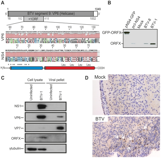
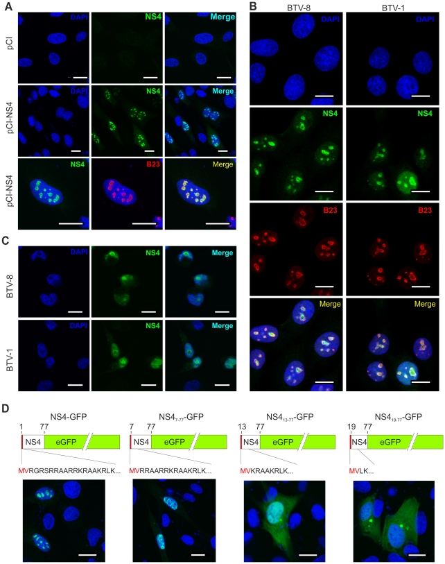
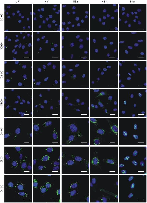
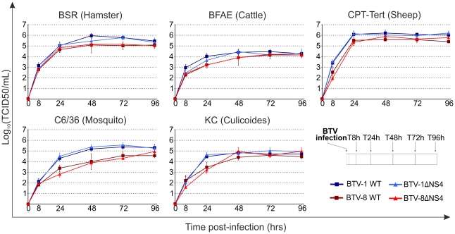
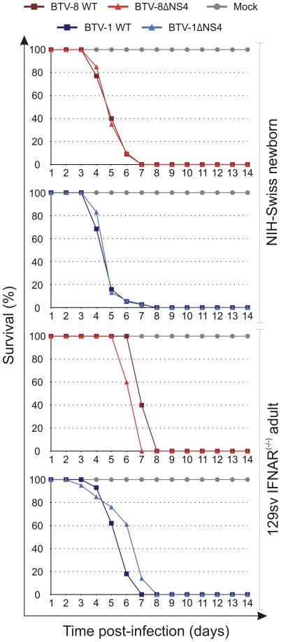
Bluetongue virus (BTV) is the causative agent of a major disease of livestock (bluetongue). For over two decades, it has been widely accepted that the 10 segments of the dsRNA genome of BTV encode for 7 structural and 3 non-structural proteins. The non-structural proteins (NS1, NS2, NS3/NS3a) play different key roles during the viral replication cycle. In this study we show that BTV expresses a fourth non-structural protein (that we designated NS4) encoded by an open reading frame in segment 9 overlapping the open reading frame encoding VP6. NS4 is 77-79 amino acid residues in length and highly conserved among several BTV serotypes/strains. NS4 was expressed early post-infection and localized in the nucleoli of BTV infected cells. By reverse genetics, we showed that NS4 is dispensable for BTV replication in vitro, both in mammalian and insect cells, and does not affect viral virulence in murine models of bluetongue infection. Interestingly, NS4 conferred a replication advantage to BTV-8, but not to BTV-1, in cells in an interferon (IFN)-induced antiviral state. However, the BTV-1 NS4 conferred a replication advantage both to a BTV-8 reassortant containing the entire segment 9 of BTV-1 and to a BTV-8 mutant with the NS4 identical to the homologous BTV-1 protein. Collectively, this study suggests that NS4 plays an important role in virus-host interaction and is one of the mechanisms played, at least by BTV-8, to counteract the antiviral response of the host. In addition, the distinct nucleolar localization of NS4, being expressed by a virus that replicates exclusively in the cytoplasm, offers new avenues to investigate the multiple roles played by the nucleolus in the biology of the cell.


Conflict of interest statement

The authors have declared that no competing interests exist.

Figures

Figure 1
Figure 1. BTV expresses a fourth non structural protein (NS4).
(A) BTV segment 9 (1049 base pairs). The VP6 protein (dark gray) is encoded by nucleotides 16 to 1002. The NS4 coding sequence is located in the +1 open reading frame (ORF) between nucleotides 182 to 418. VP6 (residues 57 to 135) and NS4 (residues 1 to 79) amino acid conservation plots are shown. NS4 secondary structure prediction indicated the presence of two putative α-helices, drawn in blue and red. The N-terminal domain (blue) is highly basic and the C-terminal domain (red) contains a conserved leucine zipper motif. (B) Western blotting of cellular extracts (lysate) of BSR cells either transfected with 1.8 µg of plasmid expressing NS4 alone (pcI-NS4) or in fusion with eGFP (peGFP-NS4), or infected by BTV-8 or BTV-1 at a MOI of 0.01. Cells were analyzed 36 h post-transfection or infection and blots were incubated with NS4 antiserum. (C) Western blots of viral pellets and cell protein extracts of BFAE cells infected by BTV-1 at a MOI of 0.05. Samples were analyzed at 48 h post-infection by SDS-PAGE and western blotting employing antisera against NS1, VP6, VP7, ORFX (NS4) and γ-tubulin as indicated. (D) Immunohistochemical detection of NS4. Immunohistochemistry was performed as described in Materials and Methods in brain tissue sections of mice inoculated with BTV-8 72 h post-infection using an antiserum against NS4. Cells expressing NS4 are stained brown as indicated by white arrows. No expression of NS4 is detected in negative control mice mock-inoculated with cell culture media.
Figure 2
Figure 2. NS4 localises in the nucleoli of transfected and infected cells.
(A) Confocal microscopy of CPT-Tert cells transfected with pCI-NS4 or empty pCI as a control. At 24 h post-transfection, cells were fixed and analyzed by immunofluorescence using antibodies against NS4 and as indicated the nucleolar marker B23 with the appropriate conjugated secondary antibodies as described in the Materials and Methods. Scale bars correspond to 18 µm. (B) Confocal microscopy of BSR cells infected by BTV-1 and BTV-8 at a MOI of 0.01. At 24 h post-infection, cells were fixed and analyzed by immunofluorescence as indicated in A. Scale bars correspond to 14 µm. (C) Confocal microscopy of C6/36 cells infected by BTV-1 and BTV-8 at a MOI of 0.05. At 48 h post-infection, cells were fixed and analyzed by immunofluorescence as for expression of NS4 as indicated in panel A. Scale bars correspond to 11 µm. (D) Confocal microscopy of CPT-Tert cells transfected with pNS4-GFP or the truncated mutants indicated above each panel. The red box corresponds to the first two amino terminal amino acid residues of NS4 that were maintained in all mutants. At 24 h post-transfection, cells were fixed and analyzed by immunofluorescence. Scale bars correspond to 18 µm.
Figure 3
Figure 3. NS4 expression profile.
Confocal microscopy of BFAE cells infected with BTV-1 at a MOI of 1.5. Cells were fixed before infection (0 h) and at 0 h30, 2 h, 4 h, 8 h, 16 h and 24 h post-infection and processed for immunofluorescence using antibodies against VP7, NS1, NS2, NS3 and NS4 with an Alexa Fluor 488 secondary antibody as described in the Materials and Methods. Scale bars correspond to 21.16 µm for 0 h to 4 h post-infection panels, and 13.6 µm for 8 h to 24 h post-infection panels.
Figure 4
Figure 4. Generation of ΔNS4 Bluetongue viruses by reverse genetics.
(A) BTV segment 9 open reading frames. VP6 amino acid residues are written in black, NS4 amino acid residues are written in grey. The nucleotides at positions 183 (T), 252 (G) and 381 (T) were mutated to C, A and A, respectively (bold). Note that whilst these mutations do not change any amino acid residues of VP6, they remove the initiation codon of NS4 (position182) and introduce two stop codons into the NS4 coding sequence at amino acid positions 24 and 67. (B) Transfected BSR cells with BTV transcripts generated in vitro (0.5×10¹¹ molecules per segment for BTV-1 and 1×10¹¹ molecules per segment for BTV-8). Cell monolayers were stained using crystal violet at 72 h post-transfection. As negative controls, ΔVP6 assays correspond to using a segment 9 containing a stop codon at position 79 in the VP6 gene. (C) Agarose gel (1.5%) of purified BTV genomic dsRNA. BSR cells infected at a MOI of 0.01 were collected at 72 h post infection and BTV dsRNA was purified as described in the Materials and Methods. 2 µg of dsRNA was loaded in each lane. (D) Western blotting of cellular extracts (lysate) of BSR cells infected at a MOI of 0.01. Cells were analyzed 36 h post-infection and blots were incubated with antisera against VP7, NS4 and γ-tubulin as indicated. Note that the double NS4 band in the BTV-1 sample is not a feature observed consistently. (E) Electron microscopy of BSR cells infected by BTV1-ΔNS4. Note cells display all the major features of BTV-infected cells including NS1 tubules (T), viral inclusion bodies (VIB) and viral particles (arrows). Scale bar = 1 µm.
Figure 5
Figure 5. In vitro growth properties of rescued WT and ΔNS4 viruses.
Growth curves of BTV-8 (dark red, square), BTV8-ΔNS4 (red, triangle), BTV-1 (blue, square) and BTV1-ΔNS4 (light blue, triangle) in cell lines derived from different species. BSR (hamster), BFAE (cattle), CPT-Tert (sheep), C6/36 (mosquito) and KC (Culicoides) cells were infected at a MOI of 0.05 and supernatants collected at 8, 24, 48, 72 and 96 h after infection. Supernatants were then titrated on BSR cells by limiting dilution analysis and the virus titers expressed as log10 (TCID50/ml). In parallel, each virus preparation was also re-titrated by limiting dilution analysis to control that equal amounts of input virus was used in each experiment. Experiments were performed independently twice, each time in duplicate, using two different virus stocks.
Figure 6
Figure 6. Cytopathic protection assay of CPT-Tert cells monolayer.
CPT-Tert cells were treated or mock treated with 1000 AVU/ml of interferon (Tau, IFNT or Universal, UIFN) for 20 h prior, and 2 h after, being infected by BTV-8 and BTV-1 (wt and ΔNS4) viruses at different MOIs (0.1, 0.01 and 0.001). Cell monolayers were stained at 72 h post-infection using crystal violet. Values indicated below each well correspond to the relative quantification (in percent) of the disrupted monolayer using Image-Pro Plus (MediaCybernetics, Inc.).
Figure 7
Figure 7. In vitro growth properties of rescued WT and ΔNS4 viruses during interferon treatment.
CPT-Tert cells were treated (solid line) or mock treated (dashed line) with 1000 AVU/ml of interferon (Tau, IFNT or Universal, UIFN) for 20 h prior and 2 h after being infected by BTV-8 (dark red, square), BTV8-ΔNS4 (red, triangle), BTV-1 (blue, square) and BTV1-ΔNS4 (light blue, triangle) viruses. Cells were infected at a MOI of 0.01. Supernatants were collected at 24, 48 and 72 h after infection, and then titrated on BSR cells by limiting dilution analysis and virus titers expressed as log10 (TCID50/ml). In parallel, each virus preparation was also re-titrated by limiting dilution analysis to control that equal amounts of input virus was used in each experiment. This experiment was performed three times, each time in duplicate.
Figure 8
Figure 8. The NS4 of BTV-1 displays similar biological properties to the homologous BTV-8 protein.
(A) Western blotting of cellular extracts (lysate) of CPT-Tert cells infected with BTV-8 wt or BTV-8ΔNS4 at a MOI of 0.01. Cells were analyzed 24 h post-infection and blots were incubated with antisera against NS1, VP7, VP6, NS4 and γ-tubulin as indicated. (B) Schematic diagram of the BTV-8/BTV-1 reassortants and mutants used in this study. Note that BTV1 and BTV8 segments/proteins are coloured in blue and red, respectively. * indicates a point mutation, while # indicates the introduction of a stop codon in the NS4 ORF. (C) CPT-Tert cells were treated with 1000 AVU/ml of Universal IFN for 20 h prior, and 2 h after, being infected by the recombinant viruses indicated in the panel using a MOI of 0.01. Cell monolayers were stained 72 h post-infection using crystal violet. Values indicated below each well correspond to the relative quantification of the disrupted monolayer using Image-Pro Plus (MediaCybernetics, Inc.). (D) CPT-Tert cells were treated (solid line) or mock treated (dashed line) with 100 AVU/ml of Universal interferon (UIFN) for 20 h prior and 2 h after being infected by the viruses indicated in the panel. Cells were infected at a MOI of 0.01. Supernatants were collected at 24, 48 and 72 h after infection, and then titrated on BSR cells by limiting dilution analysis and virus titers expressed as log10 (TCID50/ml). In parallel, each virus preparation was also re-titrated by limiting dilution analysis to control that equal amounts of input virus was used in each experiment. This experiment was performed two times, each time in duplicate.
Figure 9
Figure 9. Experimental infection of Swiss new born and IFNAR(−/−) adult mice with wt and ΔNS4 viruses.
Survival plots of either 3-days old mice inoculated intracerebrally or adult sv129 IFNAR(−/−) inoculated intraperitoneally with the following viruses: BTV-8 (dark red, square), BTV8-ΔNS4 (red, triangle), BTV-1 (blue, square) and BTV1-ΔNS4 (light blue, triangle) viruses. Mock-infected mice are shown in gray, circle. Mice were killed at two weeks post-inoculation, or earlier, if showing advanced clinical signs of systemic disease.

Similar articles

Cited by

References

    1. Mellor PS, Baylis M, Mertens PP. London: Academic Press; 2009. Bluetongue; Pastoret P-P, editor.
    1. Maclachlan NJ, Drew CP, Darpel KE, Worwa G. The pathology and pathogenesis of bluetongue. J Comp Pathol. 2009;141:1–16. - PubMed
    1. Schwartz-Cornil I, Mertens PP, Contreras V, Hemati B, Pascale F, et al. Bluetongue virus: virology, pathogenesis and immunity. Vet Res. 2008;39:46. - PubMed
    1. Erasmus BJ, Potgieter AC. The history of blueotngue. In: Mellor PS, Baylis M, Mertens P, editors. Bluetongue. Amsterdam: Elsevier; 2009. pp. 7–21.
    1. Maclachlan NJ, Guthrie AJ. Re-emergence of bluetongue, African horse sickness, and other Orbivirus diseases. Vet Res. 2010;41:35. - PMC - PubMed

Publication types

MeSH terms

Substances