

Transcription is required to establish maternal imprinting at the Prader-Willi syndrome and Angelman syndrome locus
- PMID: 22242001
- PMCID: PMC3248558
- DOI: 10.1371/journal.pgen.1002422
Transcription is required to establish maternal imprinting at the Prader-Willi syndrome and Angelman syndrome locus
Abstract
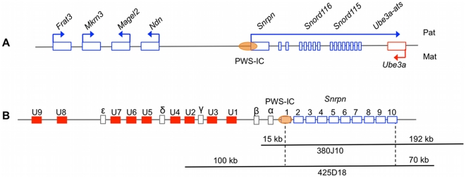
The Prader-Willi syndrome (PWS [MIM 17620]) and Angelman syndrome (AS [MIM 105830]) locus is controlled by a bipartite imprinting center (IC) consisting of the PWS-IC and the AS-IC. The most widely accepted model of IC function proposes that the PWS-IC activates gene expression from the paternal allele, while the AS-IC acts to epigenetically inactivate the PWS-IC on the maternal allele, thus silencing the paternally expressed genes. Gene order and imprinting patterns at the PWS/AS locus are well conserved from human to mouse; however, a murine AS-IC has yet to be identified. We investigated a potential regulatory role for transcription from the Snrpn alternative upstream exons in silencing the maternal allele using a murine transgene containing Snrpn and three upstream exons. This transgene displayed appropriate imprinted expression and epigenetic marks, demonstrating the presence of a functional AS-IC. Transcription of the upstream exons from the endogenous locus correlates with imprint establishment in oocytes, and this upstream exon expression pattern was conserved on the transgene. A transgene bearing targeted deletions of each of the three upstream exons exhibited loss of imprinting upon maternal transmission. These results support a model in which transcription from the Snrpn upstream exons directs the maternal imprint at the PWS-IC.
Conflict of interest statement
The authors have declared that no competing interests exist.
Figures


Similar articles
-
Prader-Willi syndrome: reflections on seminal studies and future therapies.Open Biol. 2020 Sep;10(9):200195. doi: 10.1098/rsob.200195. Epub 2020 Sep 23. Open Biol. 2020. PMID: 32961075 Free PMC article. Review.
-
Regulatory elements associated with paternally-expressed genes in the imprinted murine Angelman/Prader-Willi syndrome domain.PLoS One. 2013;8(2):e52390. doi: 10.1371/journal.pone.0052390. Epub 2013 Feb 4. PLoS One. 2013. PMID: 23390487 Free PMC article.
-
Mechanisms of activation of the paternally expressed genes by the Prader-Willi imprinting center in the Prader-Willi/Angelman syndromes domains.Proc Natl Acad Sci U S A. 2012 May 8;109(19):7403-8. doi: 10.1073/pnas.1116661109. Epub 2012 Apr 23. Proc Natl Acad Sci U S A. 2012. PMID: 22529396 Free PMC article.
-
Influence of the Prader-Willi syndrome imprinting center on the DNA methylation landscape in the mouse brain.Epigenetics. 2014 Nov;9(11):1540-56. doi: 10.4161/15592294.2014.969667. Epigenetics. 2014. PMID: 25482058 Free PMC article.
-
Genomic imprinting: potential function and mechanisms revealed by the Prader-Willi and Angelman syndromes.Mol Hum Reprod. 1997 Apr;3(4):321-32. doi: 10.1093/molehr/3.4.321. Mol Hum Reprod. 1997. PMID: 9237260 Review.
Cited by
-
RNAs of the human chromosome 15q11-q13 imprinted region.Wiley Interdiscip Rev RNA. 2013 Mar-Apr;4(2):155-66. doi: 10.1002/wrna.1150. Epub 2012 Dec 3. Wiley Interdiscip Rev RNA. 2013. PMID: 23208756 Free PMC article. Review.
-
Prader-Willi syndrome: reflections on seminal studies and future therapies.Open Biol. 2020 Sep;10(9):200195. doi: 10.1098/rsob.200195. Epub 2020 Sep 23. Open Biol. 2020. PMID: 32961075 Free PMC article. Review.
-
Maintaining memory of silencing at imprinted differentially methylated regions.Cell Mol Life Sci. 2016 May;73(9):1871-9. doi: 10.1007/s00018-016-2157-6. Epub 2016 Feb 16. Cell Mol Life Sci. 2016. PMID: 26883803 Free PMC article. Review.
-
De novo DNA methylation in the male germ line occurs by default but is excluded at sites of H3K4 methylation.Cell Rep. 2013 Jul 11;4(1):205-19. doi: 10.1016/j.celrep.2013.06.004. Epub 2013 Jun 27. Cell Rep. 2013. PMID: 23810559 Free PMC article.
-
A mouse model of Angelman syndrome imprinting defects.Hum Mol Genet. 2019 Jan 15;28(2):220-229. doi: 10.1093/hmg/ddy345. Hum Mol Genet. 2019. PMID: 30260400 Free PMC article.
References
-
- Lewis A, Reik W. How imprinting centres work. Cytogenet Genome Res. 2006;113:81–89. - PubMed
-
- Razin A, Cedar H. DNA Methylation and Genomic Imprinting. Cell. 1994;77:473–476. - PubMed
-
- Margueron R, Trojer P, Reinberg D. The key to development: interpreting the histone code? Curr Opin Genet Dev. 2005;15:163–176. - PubMed
-
- Kitsberg D, Selig S, Brandeis M, Simon I, Keshet I, et al. Allele-specific replication timing of imprinted gene regions. Nature. 1993;364:459–463. - PubMed
-
- Kishino T, Lalande M, Wagstaff J. UBE3A/E6-AP mutations cause Angelman syndrome. Nature Genetics. 1997;15:70–73. - PubMed
Publication types
MeSH terms
Substances
Grants and funding
LinkOut - more resources
Full Text Sources
Other Literature Sources
Medical
Molecular Biology Databases
Research Materials
