. 2011 Dec;7(12):e1002447.
doi: 10.1371/journal.pgen.1002447. Epub 2011 Dec 29.

DNA methylation of the gonadal aromatase (cyp19a) promoter is involved in temperature-dependent sex ratio shifts in the European sea bass

Affiliations

DNA methylation of the gonadal aromatase (cyp19a) promoter is involved in temperature-dependent sex ratio shifts in the European sea bass

Laia Navarro-Martín et al. PLoS Genet. 2011 Dec.

Abstract

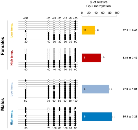
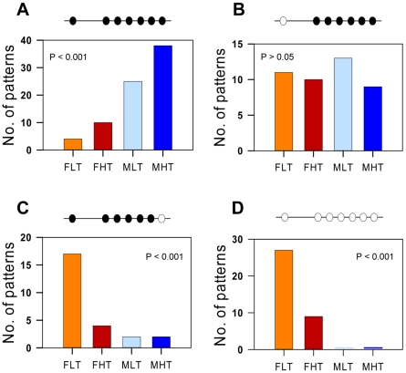
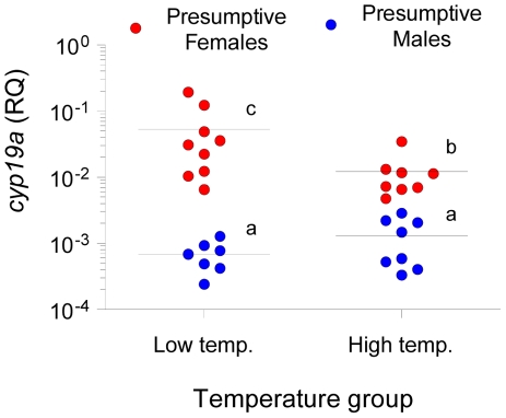
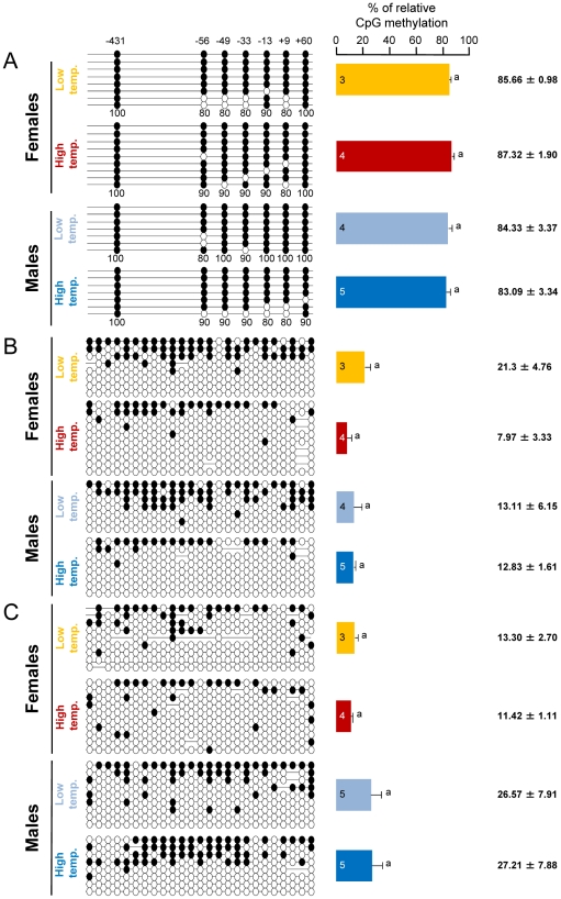
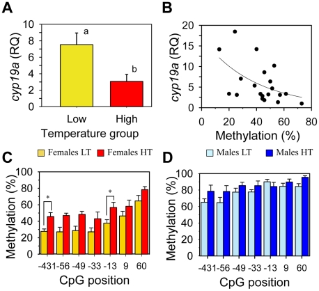
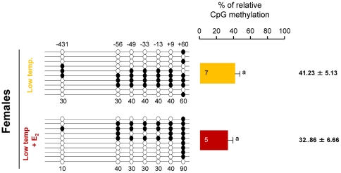
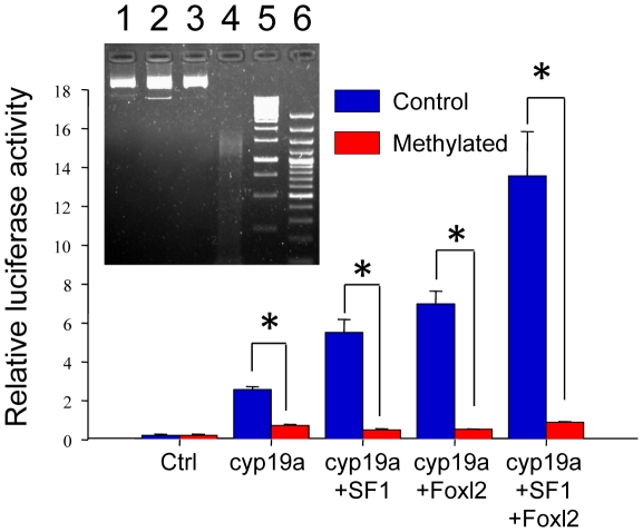
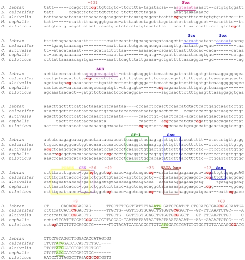
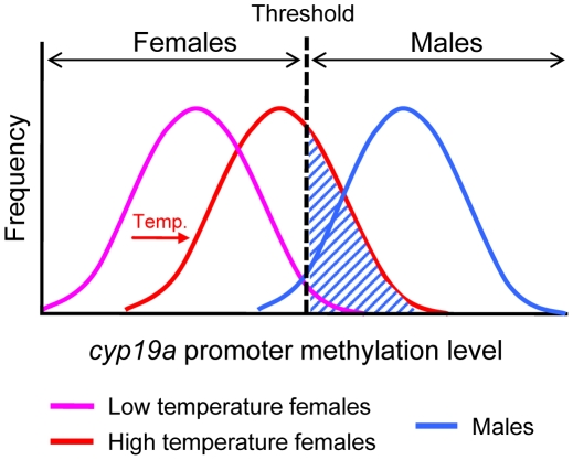
Sex ratio shifts in response to temperature are common in fish and reptiles. However, the mechanism linking temperature during early development and sex ratios has remained elusive. We show in the European sea bass (sb), a fish in which temperature effects on sex ratios are maximal before the gonads form, that juvenile males have double the DNA methylation levels of females in the promoter of gonadal aromatase (cyp19a), the enzyme that converts androgens into estrogens. Exposure to high temperature increased the cyp19a promoter methylation levels of females, indicating that induced-masculinization involves DNA methylation-mediated control of aromatase gene expression, with an observed inverse relationship between methylation levels and expression. Although different CpGs within the sb cyp19a promoter exhibited different sensitivity to temperature, we show that the increased methylation of the sb cyp19a promoter, which occurs in the gonads but not in the brain, is not a generalized effect of temperature. Importantly, these effects were also observed in sexually undifferentiated fish and were not altered by estrogen treatment. Thus, methylation of the sb cyp19a promoter is the cause of the lower expression of cyp19a in temperature-masculinized fish. In vitro, induced methylation of the sb cyp19a promoter suppressed the ability of SF-1 and Foxl2 to stimulate transcription. Finally, a CpG differentially methylated by temperature and adjacent to a Sox transcription factor binding site is conserved across species. Thus, DNA methylation of the aromatase promoter may be an essential component of the long-sought-after mechanism connecting environmental temperature and sex ratios in vertebrate species with temperature-dependent sex determination.


Conflict of interest statement

The authors have declared that no competing interests exist.

Figures

Figure 1
Figure 1. Resulting differences in sb cyp19a promoter methylation according to sex and temperature treatment.
Typical methylation patterns of sea bass gonadal aromatase promoter of one-year-old sea bass females and males that were reared at low and high temperature during early development, as observed in this study. One fish for each sex and temperature combination representative of the level of methylation is shown. Numbers with a plus or minus sign indicate CpG positions with respect to the transcription starting site. Open and filled circles denote unmethylated or methylated positions, respectively. Ten clones per fish were analyzed. Average methylation was calculated specifically for each CpG position (number below each column). The number inside the bar indicates sample size. Results as mean ± SEM. Groups with different letters are significantly different (P<0.01).
Figure 2
Figure 2. Absolute frequency of the four most frequently observed methylation patterns of the sb cyp19a promoter by sex and temperature treatment.
In all cases the expected frequency of the patterns represented here per group was >5. A, Frequency of the methylation pattern consisting of all seven positions methylated. B, Frequency of the methylation pattern with the 5′first position (−431) unmethylated and the remaining six methylated. C, Frequency of the methylation pattern with the last position (+60) unmethylated and the remaining six methylated. D, Frequency of the methylation pattern with all positions unmethylated. Abbreviations: Females at Low Temperature (FLT); Females at High Temperature (FHT); Males at Low Temperature (MLT); Males at high temperature (MHT). The level of significance is indicated for each case.
Figure 3
Figure 3. Gonadal aromatase (cyp19a) expression in sexually undifferentiated sea bass.
Gonadal aromatase expression was assessed by real time RT-PCR and used as a marker to classify fish into presumptive females (red circles) or presumptive males (blue circles) for the two thermal treatments, low and high temperature. Individual sample points are presented along with the median. Groups with different letters are significantly different (P<0.05).
Figure 4
Figure 4. Tissue and gene specificity of the effects of temperature on DNA methylation in one-year-old sea bass.
A, Resulting differences in sb cyp19a promoter DNA methylation in the brain according to sex and temperature treatment. B, Resulting differences in sb ß-actin promoter DNA methylation in the gonads according to sex and temperature treatment. C, Resulting differences in sb ß-actin promoter DNA methylation in the brain according to sex and temperature treatment. On the left, one fish representative of the level of methylation is shown per each sex and temperature combination. Open and filled circles denote unmethylated and methylated positions, respectively, while no circles denote unknown methylation status due to sequencing problems. Numbers with a plus or minus sign indicate CpG positions with respect to the transcription starting site. Average methylation was calculated for each position in each gene promoter (7 and 25 CpGs in the sb cyp19a and ß-actin gene promoters, respectively) but due to space limitations is shown only for the sb cyp19a promoter as numbers below each column (A). For each sex and temperature combination, 3–5 fish sampled at 330 dpf were used, and for each fish typically 8 clones (range 7–10) were used to determine DNA methylation levels. The number inside the bar indicates sample size. Results as mean ± SEM. Groups with the same letters are not significantly different (P>0.05).
Figure 5
Figure 5. Effects of temperature on sb cyp19a promoter methylation levels and correlation with gonadal cyp19a gene expression.
A, Female sb cyp19a expression assessed by Q-PCR; data as mean ± S.E.M (see main text for details). Groups with different letters are significantly different (P<0.01). B, Correlation between sb cyp19a methylation level and gene expression in females (male expression levels were too low to be considered). RQ, relative quantity. C and D, Differential methylation levels for each CpG position in females (C) and males (D) reared at low (LT) and high (HT) temperature; data as mean ± S.E.M. Individual CpG analysis was carried out by using Analysis of Molecular Variance (AMOVA), in which Cs were considered methylated positions and Ts unmethylated ones. Results of this analysis are summarized in Table 1.
Figure 6
Figure 6. Effects of estradiol-17ß treatment on sb cyp19a promoter methylation in one-year-old sea bass.
Result of sb cyp19a promoter methylation on untreated and treated females reared at low temperature. One fish representative of the level of methylation is shown for the treatment with or without estradiol-17ß (E2). Numbers with a plus or minus sign indicate CpG positions with respect to the transcription starting site. Open and filled circles denote unmethylated and methylated positions, respectively. Ten clones per fish were analyzed. Average methylation was calculated specifically for each position (number below each column). The number inside the bar indicates sample size. Results as mean ± SEM. Groups with the same letter are not significantly different (P>0.05).
Figure 7
Figure 7. Effects of methylation on sea bass cyp19a promoter activity in vitro.
HEK293T cells were transfected with pGL3-cyp19a methylated and unmethylated promoter vectors. Transcription factors SF1 and Foxl2 were cotransfected with the sb cyp19a promoter to activate promoter luciferase activity. Transfected methylated and unmethylated groups were as follows (see Materials and Methods for amounts used): 1) plasmid β-galactosidase (Ctrl); 2) sb cyp19a promoter cloned into pGL3-basic luciferase reporter plasmid (cyp19a); 3) sb cyp19a and tilapia SF1 cloned into pCDNA3.1 expression plasmid (cyp19a+SF1); 4) sb cyp19a and tilapia Foxl2 cloned into pCDNA3.1 expression plasmid (cyp19a+Foxl2); 5) sb cyp19a, tilapia SF1 and Foxl2 cloned into pCDNA3.1 (cyp19a+SF1+Foxl2). The Student's t-test was used to compare methylated and unmethylated vectors. Significant differences are denoted by an asterisk and were as follows: cyp19a, P = 0.006; cyp19a+SF1, P = 0.006; cyp19a+Foxl2, P = 0.003; cyp19a+SF1+Foxl2, P = 0.013. Also, SF1 and Foxl2 exhibited an additive effect since the activation of sb cyp19a promoter was significantly higher when both were transfected together. Values are shown as mean ± S.E.M. (n = 2–4). Insert: Successful vector methylation verification by analysis of band patterns on electrophoresis gel after digestion of the purified plasmids with the McrBC enzyme. Lane 1, 0.5 µg sb cyp19a-pGL3; lane 2, 0.5 µg sb cyp19a-pGL3 treated with McrBC; lane 3, 0.5 µg SssI-methylated sb cyp19a-pGL3; lane 4, 0.5 µg SssI-methylated sb cyp19a-pGL3 treated with McrBC; lane 5, 1 Kb marker; lane 6, 100 bp marker. The electrophoresis gel shows that, as expected, only the methylated vector was digested.
Figure 8
Figure 8. Alignment of the sea bass gonadal cyp19a promoter with that of other teleost species.
Accession numbers: Dicentrarchus labrax, DQ177458; Lates calcarifer, AY686690; Cromileptes altivelis, AY686691; Mugil cephalus, AY859426; Oerochromis niloticus, AB089924. CpG dinucleotides are shown in bold. The numbers above D. labrax CpGs denote the position relative to the transcription start site. Putative binding sites for specific transcription factors were localized in D. labrax after Galay-Burgos et al. and are indicated by boxes when are conserved among species or by underlying if apply only to D. labrax. The open reading frame is depicted by upper case text and putative transcription start sites “ATG” are highlighted in bold and underlined.
Figure 9
Figure 9. Schematic diagram of the frequency distribution of the sb cyp19a promoter methylation levels in females and males.
Low methylation levels (purple curve) of the sb cyp19a promoter are typical of females and are required for ovarian differentiation. Males typically show high methylation levels (blue curve) of the cyp19a promoter. This blocks aromatase transcription and induces testicular differentiation caused by lack of estrogens. As shown in this study, high temperatures (HT) during the thermosensitive period increase methylation levels of the sb cyp19a promoter (red curve), shifting the female distribution closer to that of males. The dashed area represents the fraction of genotypic females that are sex reversed into phenotypic males by high temperature.

Similar articles

Cited by

References

    1. Valenzuela N, Lance V. Temperature-dependent sex determination in vertebrates. Washington: Smithsonian Books; 2004.
    1. Pieau C, Dorizzi M. Oestrogens and temperature-dependent sex determination in reptiles: all is in the gonads. Journal of Endocrinology. 2004;181:367–377. - PubMed
    1. Ramsey M, Crews D. Steroid signaling and temperature-dependent sex determination. Reviewing the evidence for early action of estrogen during ovarian determination in turtles. Seminars in Cell & Developmental Biology. 2009;20:283–292. - PMC - PubMed
    1. Ospina-Álvarez N, Piferrer F. Temperature-dependent sex determination in Fish. Prevalence, existence of a single sex ratio response pattern, and possible effects on climate change. Public Library of Science One. 2008;3:e2837. - PMC - PubMed
    1. Van Nes S, Andersen O. Temperature effects on sex determination and ontogenetic gene expression of the aromatases cyp19a and cyp19b, and the estrogen receptors esr1 and esr2 in Atlantic halibut (Hippoglossus hippoglossus). Molecular Reproduction and Development. 2006;73:1481–1490. - PubMed

Publication types