. 2012 Jan;8(1):e1002425.
doi: 10.1371/journal.pgen.1002425. Epub 2012 Jan 5.

Microenvironmental regulation by fibrillin-1

Affiliations

Microenvironmental regulation by fibrillin-1

Gerhard Sengle et al. PLoS Genet. 2012 Jan.

Abstract

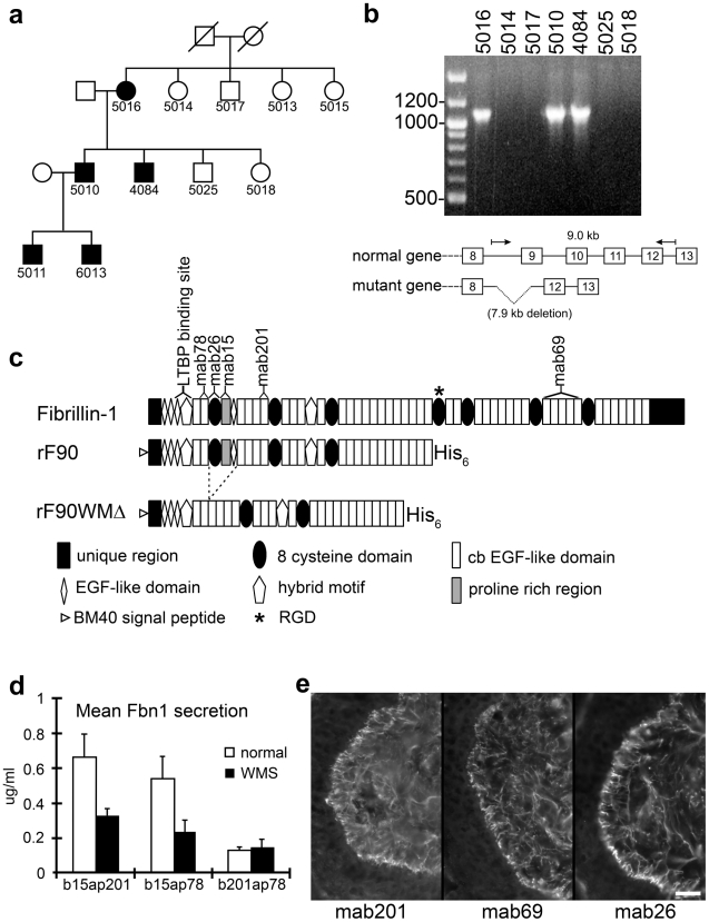
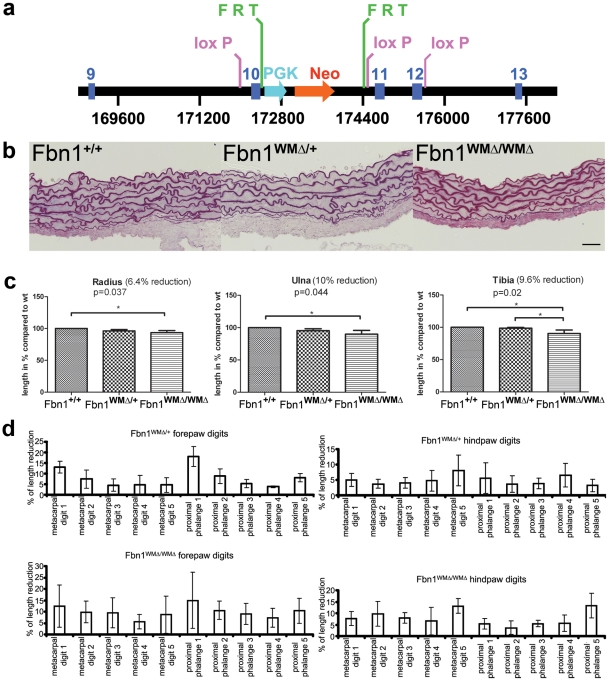
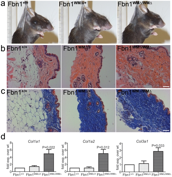
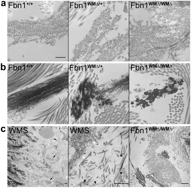
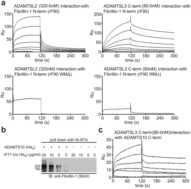
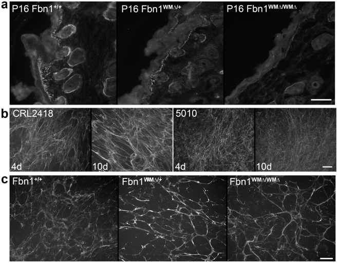
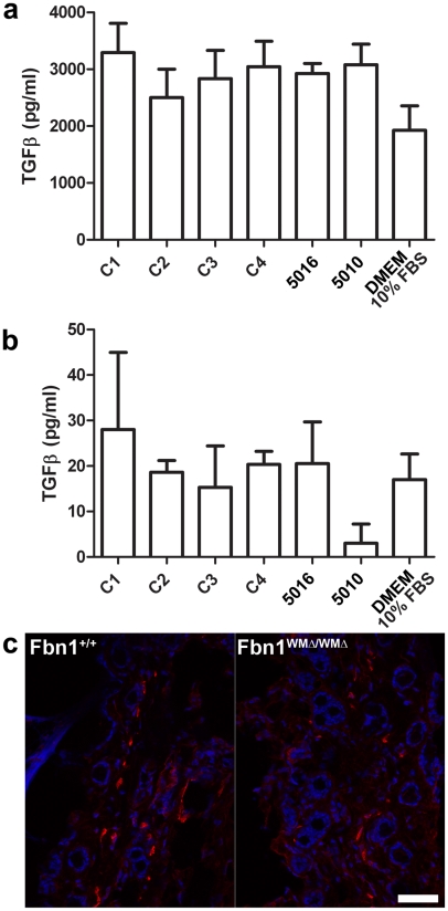
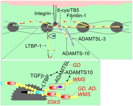
Fibrillin-1 is a ubiquitous extracellular matrix molecule that sequesters latent growth factor complexes. A role for fibrillin-1 in specifying tissue microenvironments has not been elucidated, even though the concept that fibrillin-1 provides extracellular control of growth factor signaling is currently appreciated. Mutations in FBN1 are mainly responsible for the Marfan syndrome (MFS), recognized by its pleiotropic clinical features including tall stature and arachnodactyly, aortic dilatation and dissection, and ectopia lentis. Each of the many different mutations in FBN1 known to cause MFS must lead to similar clinical features through common mechanisms, proceeding principally through the activation of TGFβ signaling. Here we show that a novel FBN1 mutation in a family with Weill-Marchesani syndrome (WMS) causes thick skin, short stature, and brachydactyly when replicated in mice. WMS mice confirm that this mutation does not cause MFS. The mutation deletes three domains in fibrillin-1, abolishing a binding site utilized by ADAMTSLIKE-2, -3, -6, and papilin. Our results place these ADAMTSLIKE proteins in a molecular pathway involving fibrillin-1 and ADAMTS-10. Investigations of microfibril ultrastructure in WMS humans and mice demonstrate that modulation of the fibrillin microfibril scaffold can influence local tissue microenvironments and link fibrillin-1 function to skin homeostasis and the regulation of dermal collagen production. Hence, pathogenetic mechanisms caused by dysregulated WMS microenvironments diverge from Marfan pathogenetic mechanisms, which lead to broad activation of TGFβ signaling in multiple tissues. We conclude that local tissue-specific microenvironments, affected in WMS, are maintained by a fibrillin-1 microfibril scaffold, modulated by ADAMTSLIKE proteins in concert with ADAMTS enzymes.


Conflict of interest statement

The authors have declared that no competing interests exist.

Figures

Figure 1
Figure 1. Characterization of a novel genomic deletion in FBN1 in a family with WMS.
(a) Partial pedigree of the family. Affected individuals are shown as filled symbols. (b) PCR from genomic DNA of selected family members, using primers flanking the deleted region. Only the affected individuals give rise to the shortened product (1.1 kb), which after DNA sequencing revealed a 7895 nt deletion with boundaries in FBN1 introns 8 (IVS8-1207) and 11 (IVS11+1257). The wildtype product (9.0 kb) was too large to be amplified under the conditions used. (c) Schematic drawing of domains contained in the recombinant N-terminal half of wildtype (rF90) and WMS deleted fibrillin-1 protein (rF90WMΔ). The mutation results in the deletion of exons 9–11, encoding the first 8-cysteine domain, the adjacent proline rich region, and a generic EGF-like domain. Purity of preparations of rF90 and rF90WMΔ is shown in Figure S3. Epitopes of monoclonal antibodies used in this study are shown above the full-length molecule. In addition, the single RGD site and the binding site for LTBP are marked. (d) Sandwich ELISA used to quantitate secretion of wildtype and mutant fibrillin-1 in culture medium from normal dermal fibroblasts and WMS fibroblasts. Capture antibodies were biotinylated (b), and detector antibodies were coupled to alkaline phosphatase (ap). The capture and detector antibody pair that recognizes epitopes outside of the deleted region (b201 and ap78) detected both normal and WMS mutant fibrillin molecules and showed equal levels of fibrillin-1 secretion in both wildtype and WMS fibroblast medium. When mAb15 was used as a capture antibody, only wildtype fibrillin-1 was detected, because the proline-rich region, which contains the epitope recognized by mAb15 is deleted in WMS fibrillin-1. Quantitation of fibrillin-1 secretion using the b15ap201 and b15ap78 antibody pairs showed approximately half the amount of fibrillin-1 in WMS fibroblast medium compared to medium from normal fibroblasts. These results indicate that equal amounts of normal and mutated fibrillin-1 are secreted by WMS fibroblasts. The differences in total fibrillin-1 amounts measured using the different pairs are due to differences in affinities of the pairs for the protein standard (rF11). For this experiment, n = 3 and the error bars represent the standard deviation. (e) Immunofluorescence of WMS skin showed a normal fibrillin pattern when mAb201, mAb69, or mab26 were used. Unlike immunofluorescence of Marfan skin , fibrillin-1 staining patterns were not fragmented in WMS skin. Scale bar = 20 µm.
Figure 2
Figure 2. Replication of the WMS mutation in mice.
(a) The targeted Fbn1 locus. A neomycin selection cassette (PGK-Neo, flanked by FRT sites) was placed in the intron between exons 10 and 11. In addition, loxP sites were introduced before exons 10 and 11 and after exon 12. The neomycin cassette was removed by breeding targeted mice to FLPe mice. Cre-mediated recombination of the loxP sites resulted in deletion of exons 10–12, replicating the human WMS mutation. (b) Aortic root morphology. In contrast to Marfan mice, the aortic roots of 10 month old mutant mice showed no signs of fragmentation of the elastic lamellae. Scale bar = 25 µm. (c) Length measurements of long bones. μCT measurements revealed a reduction of 6–10% at 1 month of age, when homozygous mice were compared to gender-matched wildtype littermates (each bar represents the mean and standard deviation of measurements from 4 animals, n = 4). Significant p-values were obtained for all bones when comparisons were between homozygous and wildtype littermates. (d) Length measurements of skeletal elements in forepaws and hindpaws showed reduced digit length of metacarpals and proximal phalanges in WMΔ mutant mice relative to wildtype mice. All analyzed animals were gender matched littermates at 1 month of age (n = 5 for each genotype).
Figure 3
Figure 3. Thick skin phenotype in WMΔ mice.
(a) Gross inspection. 7.5 month WMΔ mice could be identified by touch. Skin felt thicker and was less elastic in both heterozygous (WMΔ/+) and homozygous (WMΔ/WMΔ) mice compared to wildtype (Fbn1+/+) littermates. Here, mice were sacrificed, shaved, and immediately suspended by forceps positioned at the same relative spot between the ears. The ruler is included to show equivalent magnifications in the photographs. (b) Hematoxylin and eosin staining of skin from 10 month old WMΔ littermates. Dermal fibers in the mutant skin appeared to be thicker and more densely packed when compared to the wildtype littermate skin. Scale bar = 50 µm. (c) Masson's trichrome staining of skin from the same 10 month old littermates as in (b). WMΔ heterozygous and homozygous mutant skin showed increased collagen deposition. In addition, in both (b) and (c), the dermis appeared to be wider with a strikingly diminished hypodermal fat layer. Scale bar = 50 µm. (d) qPCR of collagen genes. RNAs were extracted from skin from WMΔ wildtype and mutant littermates (n = 5 for each genotype), and collagen genes were quantitated by qPCR. Type I and Type III collagen gene expression was found to be significantly upregulated in the homozygotes.
Figure 4
Figure 4. Ultrastructural abnormalities in microfibrils in WMΔ mouse and WMS human skin.
(a,b) Immunogold labeling of 9 month old wildtype and WMΔ mutant skin. Wildtype microfibrils decorated with fibrillin-1 antibodies showed periodic gold labeling along the lengths of individual microfibrils in the absence of elastin (Fbn1+/+, a) and on the periphery of amorphous, darkly stained elastin cores (Fbn1+/+, b). In contrast, fibrillin microfibrils in WMΔ mutant skin showed reduced immunogold periodicity along microfibrils and larger and denser accumulations of microfibril aggregates (WMΔ/+ and WMΔ/WMΔ, a); elastic fibers appeared to be moth-eaten and also showed reduced fibrillin-1 periodic labeling (b). Scale bar = 300 nm for all panels in a,b. (c) Extreme ultrastructural appearance of abnormal microfibril aggregates in both human WMS and mouse WMΔ skin. Large dense aggregates of microfibrils were easily identified at low magnification in the skin of an 18 year old individual with WMS (left panel, arrows). Immunogold labeling demonstrated both normal periodic microfibrils (black arrowheads) and irregularly labeled microfibrils (white arrowheads) (middle panel). Large dense aggregates of microfibrils were also found in older WMΔ mutant mouse skin. One such aggregate is shown in skin from a 17 month old homozygous (WMΔ/WMΔ) mouse. Scale bars = 500 nm.
Figure 5
Figure 5. Biochemical analyses of interactions among ADAMTSL proteins, fibrillin-1, and ADAMTS-10.
(a) SPR sensorgrams showing binding of different concentrations of soluble ligands to the N-terminal half of fibrillin-1 (rF90), coupled onto a chip. Full-length ADAMTSL-2 (320-0 nM) interacts with rF90, as does the C-terminal end of ADAMTSL-3 (80-0 nM). No binding was detected when rF90WMΔ was used, demonstrating that the binding site for ADAMTSL proteins resides in the deleted region. (b) Fibrillin-1/ADAMTS-10 pull-down assay. Conditioned medium from transfected cells expressing full-length ADAMTS-10 with a C-terminal His6-tag was incubated with increasing amounts of rF11 (N-terminal half of fibrillin-1, similar to rF90 but lacking the His6-tag). ADAMTS-10 complexes were pulled down after incubation with Nickel NTA resin. SDS-PAGE followed by immunoblotting (IB) with anti-fibrillin-1 antibody (9543) showed the presence of fibrillin-1 in the pulled-down ADAMTS-10 complexes. Conditioned medium from untransfected cells (−) was subjected to the same procedure and served as a control. Even though rF11 was added in similar amounts to the control medium, no fibrillin-1 was pulled-down, demonstrating the specificity of the interaction between ADAMTS-10 and fibrillin-1. (c) SPR sensorgrams showing binding of different concentrations (80-0 nM) of soluble C-terminal ADAMTSL-3 to the C-terminal end of ADAMTS-10, coupled to a chip. Calculated KD for this interaction was 2 nM.
Figure 6
Figure 6. Immunofluorescence analyses of WMS skin and fibroblast cultures.
(a) Immunolocalization of ADAMTSL-6 on P16 wildtype, heterozygous and homozygous WMΔ littermates. Antibodies specific for ADAMTSL-6 were used to stain littermate skin. As previously shown , ADAMTSL-6 staining was similar to fibrillin-1 immunostaining in wildtype skin. WMΔ mutant skin showed a reduction in ADAMTSL-6 immunostaining, indicating that deletion of the ADAMTSL binding site in fibrillin-1 prevents ADAMTSL-6 from binding to fibrillin-1 in vivo. (b) Human fibroblast cell cultures stained with antibodies to fibrillin-1 (pAb9543) after 4 days and 10 days of culture. Control (CRL2418) fibroblasts elaborated a typical fibrillin-1 fibril matrix that was clearly abundant at day 4 and day 10. In contrast, WMS (5010) fibroblasts deposited abundant fibrillin-1 fibrils at day 4, but these fibrils were thinner and more diffuse. Even after 10 days in culture, the WMS fibroblasts failed to establish prominent bundles of fibrillin fibrils. (c) Fibroblast cultures from wildtype, heterozygous, and homozygous WMΔ mice stained with anti-fibrillin-1 (pAb9543) after 4 days in culture. Mutant fibroblast cultures elaborated typical abundant, long fibrillin-1 fibrils that appeared somewhat thicker than fibrils in wildtype cultures. Scale bars = 50 µm.
Figure 7
Figure 7. Measurements of TGF-β in cultured fibroblasts; α-smooth muscle actin staining.
(a) WMS fibroblasts (family members 5016 and 5010) secreted equal amounts of total TGF-β protein compared to controls (C1, C2, C3, and C4). (b) No significant differences were detected in amounts of active TGF-β present in the media of WMS fibroblasts compared to controls. Medium containing 10% fetal bovine serum was used to show baseline values. For experiments in (a) and (b), n = 2 or 3, and the error bars represent the standard deviation. (c) Skin from 6-month old wildtype and WMΔ/WMΔ littermates showed no difference in numbers of cells stained by an α-smooth muscle actin antibody (red). DAPI nuclear stain is blue. Scale bar = 20 µm.
Figure 8
Figure 8. Model of fibrillin-1 containing microfibrils showing the locations of binding sites for ADAMTSL proteins, LTBP-1, and integrins.
This model of fibrillin-1 molecules arranged as parallel, staggered molecules within the beads-on-a-string microfibril was previously proposed . Two staggered fibrillin-1 molecules are shown with colored domains (see Figure 1c for domain structure), while other fibrillin molecules within the microfibril are depicted as dashed black lines. Beaded regions of the microfibril are represented as gray scalloped circles. The inset shows the N-terminus (black) of one molecule extending through cbEGF5 and crossing over the middle portion of a second molecule (shown from Hybrid2 through cbEGF27). In this model, binding sites for ADAMTSL proteins (within the first 8-cysteine domain, the proline-rich domain, and the adjacent generic EGF-like domain) and for LTBP-1 (within the first hybrid domain) on one molecule are very close to the integrin-binding RGD site (contained in the fourth 8-cysteine domain) on a second molecule. Mutations in the fourth 8-cysteine domain can cause SSKS, presumably by disrupting integrin binding. The fifth 8-cysteine domain or TB5 contains mutations in FBN1 that result in WMS , geleophysic (GD) or acromicric dysplasia (AD) . Mutations in ADAMTSL2 also lead to GD, and mutations in ADAMTS10 lead to WMS. We propose that this cluster of molecular interactions (magnified in the inset) constitutes a microenvironment controlling thick skin and musculoskeletal growth.

Similar articles

Cited by

References

    1. McKusick VA. The Weill-Marchesani syndrome. In: McKusick VA, editor. Heritable disorders of connective tissue. 4th edition. St. Louis, MO: CV Mosby Company; 1972. pp. 282–291.
    1. Wirtz MK, Samples JR, Kramer PL, Rust K, Yount J, et al. Weill-Marchesani syndrome–possible linkage of the autosomal dominant form to 15q21.1. Am J Med Genet. 1996;65:68–75. - PubMed
    1. Faivre L, Gorlin RJ, Wirtz MK, Godfrey M, Dagonneau N, et al. In frame fibrillin-1 gene deletion in autosomal dominant Weill-Marchesani syndrome. J Med Genet. 2003;40:34–36. - PMC - PubMed
    1. Faivre L, Megarbane A, Alswaid A, Zylberbeg L, Aldohayan N, et al. Homozygosity mapping of a Weill-Marchesani syndrome locus to chromosome 19p13.3-p13.2. Hum Genet. 2002;110:366–370. - PubMed
    1. Dagoneau N, Benoist-Lasselin C, Huber C, Faivre L, Megarbane A, et al. ADAMTS10 mutations in autosomal recessive Weill-Marchesani syndrome. Am J Hum Genet. 2004;75:801–806. - PMC - PubMed

Publication types

MeSH terms

LinkOut - more resources