. 2011;6(12):e29498.
doi: 10.1371/journal.pone.0029498. Epub 2011 Dec 29.

Stress-induced C/EBP homology protein (CHOP) represses MyoD transcription to delay myoblast differentiation

Affiliations

Stress-induced C/EBP homology protein (CHOP) represses MyoD transcription to delay myoblast differentiation

Joel Alter et al. PLoS One. 2011.

Abstract

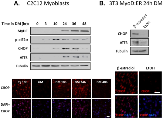
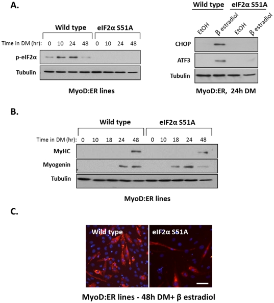
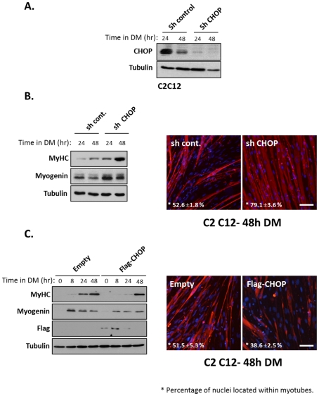
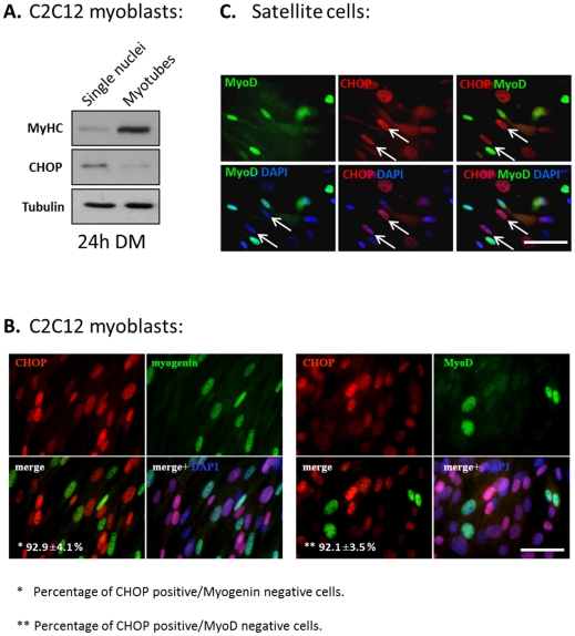
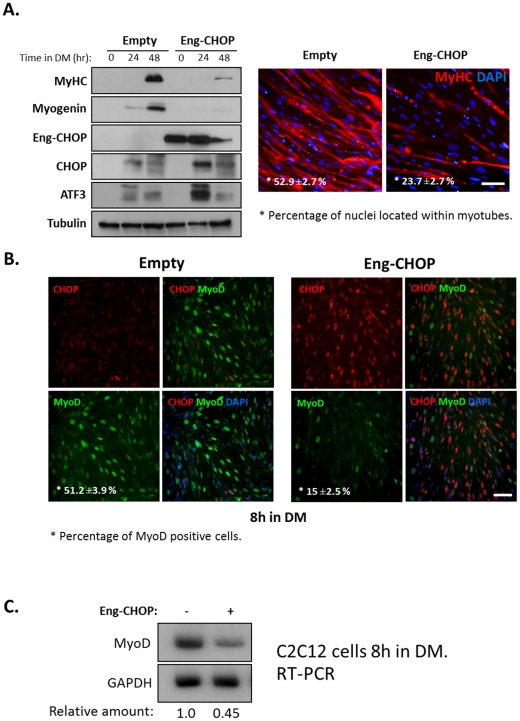
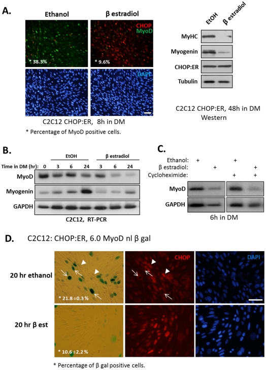
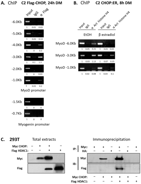
When mouse myoblasts or satellite cells differentiate in culture, the expression of myogenic regulatory factor, MyoD, is downregulated in a subset of cells that do not differentiate. The mechanism involved in the repression of MyoD expression remains largely unknown. Here we report that a stress-response pathway repressing MyoD transcription is transiently activated in mouse-derived C2C12 myoblasts growing under differentiation-promoting conditions. We show that phosphorylation of the α subunit of the translation initiation factor 2 (eIF2α) is followed by expression of C/EBP homology protein (CHOP) in some myoblasts. ShRNA-driven knockdown of CHOP expression caused earlier and more robust differentiation, whereas its constitutive expression delayed differentiation relative to wild type myoblasts. Cells expressing CHOP did not express the myogenic regulatory factors MyoD and myogenin. These results indicated that CHOP directly repressed the transcription of the MyoD gene. In support of this view, CHOP associated with upstream regulatory region of the MyoD gene and its activity reduced histone acetylation at the enhancer region of MyoD. CHOP interacted with histone deacetylase 1 (HDAC1) in cells. This protein complex may reduce histone acetylation when bound to MyoD regulatory regions. Overall, our results suggest that the activation of a stress pathway in myoblasts transiently downregulate the myogenic program.


Conflict of interest statement

Competing Interests: The authors have declared that no competing interests exist.

Figures

Figure 1
Figure 1. Transient expression of stress response proteins during myoblast differentiation.
(A) C2C12 myoblasts were differentiated in DM and proteins were extracted at different time points as indicated. Protein were separated and analyzed by Western blotting (Upper panel). In another experiment C2C12 myoblasts were differentiated as is described. Cells were fixed and analyzed by immunostaining with an antibody to CHOP. CHOP in red, DAPI in blue (Lower panel). Bar, 50 µm. (B) 3T3 MyoD:ER cells were differentiated in DM containing ethanol or β estradiol (0.1 µM) for 24 hours. Proteins were extracted and were analyzed by Western blot with the indicated antibodies (Upper panel). Cells were fixed and were analyzed by immunostaining with an antibody to CHOP. CHOP is red, DAPI in blue (Lower panel). Bar, 50 µm.
Figure 2
Figure 2. Muscle differentiation of eIF2αS51A knockin cells.
Wild type eIF2α and mutated eIF2αS51A fibroblasts were infected with viruses encoding MyoD:ER protein. (A) Cells were allowed to differentiate in DM and β estradiol (0.1 µM) for the indicated time periods and proteins were analyzed by Western blot (left panel). Cells were grown in DM and ethanol or β estradiol (0.1 µM) for 24 hours and CHOP and ATF3 proteins were analyzed by Western blot (right panel). (B) Cell lines were grown as is described in A, and were analyzed by Western blot. (C) Cell lines were grown in DM for 48 hours. Cells were immunostained with an anti MyHC antibody (MF20); MyHC in red, DAPI in blue. Bar, 50 µm.
Figure 3
Figure 3. CHOP inhibits myogenic differentiation.
(A) CHOP was knockdown in C2C12 myoblasts by infection of lentivirus expressing ShRNA. The levels of CHOP protein were analyzed by Western blot of infected myoblasts. (B) Infected myoblasts were grown in DM for the indicated time periods and myogenic markers were analyzed by Western blot (left panel). Infected myoblasts were grown in DM for 48 hours before cells were immunostained with anti MyHC antibodies (MF20) (right panel) MyHC in red, DAPI in blue. Percentage of nuclei in myotubes was calculated from three independent experiments. Mean values and standard errors are presented. Bar, 50 µm. (C) C2C12 myoblasts were infected with retroviruses encoding a flag-tagged CHOP protein or the parental retrovirus serving as a control. Infected myoblasts were grown in DM for the indicated time periods and myogenic markers were analyzed by Western blot (left panel). Infected myoblasts were grown in DM for 48 hours before cells were immunostained with anti MyHC antibodies (MF20) (right panel) MyHC in red, DAPI in blue. Percentage of nuclei in myotubes was calculated from three independent experiments. Mean values and standard errors are presented. Bar, 50 µm.
Figure 4
Figure 4. The expression of CHOP and MRFs is mutual exclusive.
(A) C2C12 cells were grown in DM for 24 hours and mononucleated cells were separated from myotubes by selective trypsinization. The two cell populations were subjected to Western blot for analyzing the expression of CHOP. (B) C2C12 myoblasts were grown in DM for 24 hours, and double stained with antibodies directed against CHOP and myogenin (left panel) or with antibodies directed against CHOP and MyoD (right panel). DAPI in blue, MyoD and myogenin in green and CHOP in red. Percentage of CHOP positive, myogenin negative and CHOP positive, MyoD negative relative to the total number of CHOP positive cells was calculated in three independent experiments. Mean values and standard errors are presented. Bar, 50 µm. (C) The expression of CHOP in primary satellite cells. To induce their differentiation, satellite cells were grown for 24 hours in GM medium. Cells were analyzed by immunostaining with anti-MyoD (green) and anti-CHOP (red) antibodies. DAPI staining is in blue. Arrows point at nuclei positive for CHOP staining and negative for MyoD staining. Bar, 50 µm.
Figure 5
Figure 5. CHOP functions a transcription repressor in myoblasts.
(A) A retrovirus encoding a chimera Engrailed-CHOP protein or a retrovirus containing the parental vector was used to infect C2C12 myoblasts. Infected cells were grown in GM and in DM for the indicated time periods and proteins were analyzed by Western blot (left panel). Infected myoblasts were grown in DM for 48 hours and were immunostained with an anti-MyHC antibody (MF20) (right panel). MyHC staining is in red and DAPI is in blue. Percentage of nuclei in myotubes was calculated from three independent experiments. Mean values and standard errors are presented. Bar, 50 µm. (B) Infected cells described in A were grown in DM for 8 hours and were analyzed by immunostaining with antibodies directed against CHOP and MyoD. Control infected cells (left panel) and Engrailed-CHOP infected cells (right panel). Percentage of MyoD-positive nuclei relative to the total number of nuclei was calculated in three independent experiments. Mean values and standard errors are presented. Bar, 50 µm. (C) C2C12 infected cells as in A were grown in DM for 8 hours and total RNA was then extracted. MyoD mRNA levels were analyzed by semi-quantitative RT-PCR and quantified by the posphoimager.
Figure 6
Figure 6. CHOP represses MyoD transcription.
(A) A C2C12 derived cell line expressing a chimera CHOP:ER protein was constructed as is described under “Materials and Methods”. (A) Myoblasts were grown in the presence of ethanol or β estradiol (0.1 µM) for 8 hours. Cells were immunostained using anti-MyoD and anti-CHOP antibodies. DAPI in blue, MyoD in green and CHOP in red. Percentage of MyoD-positive nuclei relative to the total number of nuclei was calculated. Bar, 50 µm. (left panel). In another experiment, cells were grown in the presence of ethanol or β estradiol (0.1 µM) for 48 hours and proteins were analyzed by Western blot (right panel). (B) The same cells as above were grown in DM in the presence of ethanol or β estradiol (0.1 µM) for the indicated time periods and mRNA levels of myod and myogenin were determined by semi-quantitative RT-PCR analysis. (C) The same cells as above were grown in DM and in the presence of ethanol or β estradiol for 6 hours in the absence or presence of cycloheximide added to cells one hour before the addition of ethanol or β estradiol. (D) A clone of the above cells (i.e., expressing CHOP:ER) with integrated MyoD reporter gene (6.0 MyoD -nl β Gal) was isolated. These cells were grown in the presence of ethanol or β estradiol for 20 hours. Nuclear expression of β Gal was identified by an enzymatic colorimetric assay, and the expression of CHOP by immunostaining. Arrows point at β Gal-positive nuclei that are CHOP negative. Percentage of β Gal-positive nuclei out of the total number of nuclei was calculated in two independent experiments. Mean values and standard errors are presented. Bar, 50 µm.
Figure 7
Figure 7. CHOP associates with MyoD regulatory sequences and affects histone acetylation.
(A) A Chromatin IP experiment was performed on C2C12 cells expressing Flag-CHOP that were grown in DM for 24 hours. Immunoprecipitation of fragmented DNA was performed with anti-Flag antibodies. PCR amplification of fragments scattered throughout 6 Kb upstream region of the MyoD transcription unit and along 2 Kb upstream of the myogenin transcription unit was performed. PCR fragments were separated over agarose gels. Gels were scanned and values of band intensities are presented below. For each set of PCR primers, the value of the input was set to 1. (B) C2C12 cells expressing CHOP:ER chimera were grown in DM for 8 hours in the presence of ethanol or β estradiol (0.1 µM). Chromatin IP assay was performed on fragmented DNA with anti-acetylated histone H4 antibody. PCR amplification of fragments scattered along 6 Kb upstream of the MyoD transcription unit was performed. Gels were scanned and values of band intensities are presented below. For each set of PCR primers, the value of the input was set to 1 (C) 293T cells were transfected with expression plasmids as indicated. Cells were lysed under mild conditions and extracted proteins were analyzed (left) or immunoprecipitated with anti-Myc or anti-HA epitope antibodies as indicated (right). Proteins were analyzed by Western blotting as indicated.

References

    1. Tajbakhsh S. Skeletal muscle stem cells in developmental versus regenerative myogenesis. J Intern Med. 2009;266:372–389. - PubMed
    1. Beauchamp JR, Heslop L, Yu DS, Tajbakhsh S, Kelly RG, et al. Expression of CD34 and Myf5 defines the majority of quiescent adult skeletal muscle satellite cells. J Cell Biol. 2000;151:1221–1234. - PMC - PubMed
    1. Fuchtbauer EM, Westphal H. MyoD and myogenin are coexpressed in regenerating skeletal muscle of the mouse. Dev Dyn. 1992;193:34–39. - PubMed
    1. Kitzmann M, Carnac G, Vandromme M, Primig M, Lamb NJ, et al. The muscle regulatory factors MyoD and myf-5 undergo distinct cell cycle-specific expression in muscle cells. J Cell Biol. 1998;142:1447–1459. - PMC - PubMed
    1. Megeney LA, Kablar B, Garrett K, Anderson JE, Rudnicki MA. MyoD is required for myogenic stem cell function in adult skeletal muscle. Genes Dev. 1996;10:1173–1183. - PubMed

Publication types

MeSH terms