. 2012 Jan 24;51(3):776-85.
doi: 10.1021/bi2017427. Epub 2012 Jan 12.

Probing structural features of Alzheimer's amyloid-β pores in bilayers using site-specific amino acid substitutions

Affiliations

Probing structural features of Alzheimer's amyloid-β pores in bilayers using site-specific amino acid substitutions

Ricardo Capone et al. Biochemistry. .

Abstract

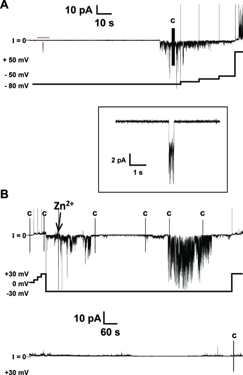
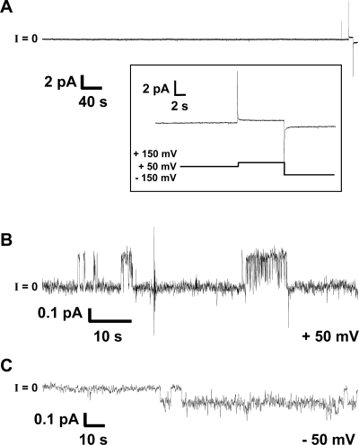
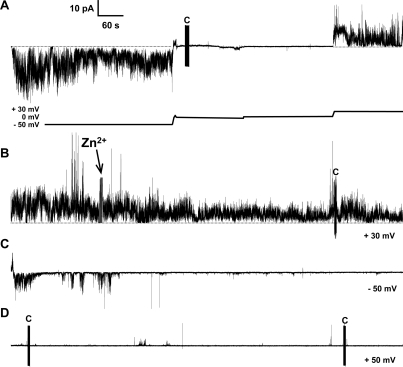
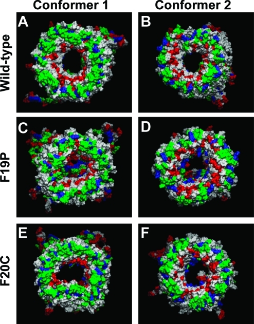
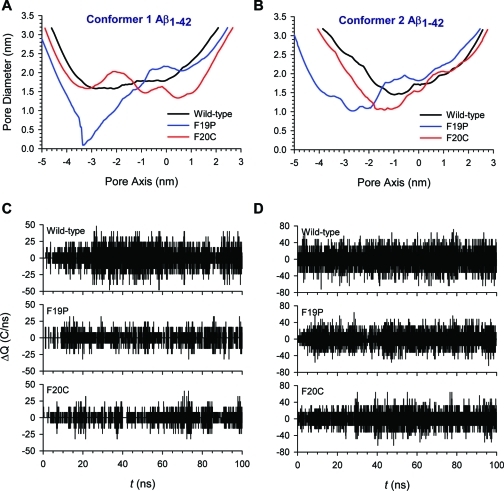
A current hypothesis for the pathology of Alzheimer's disease (AD) proposes that amyloid-β (Aβ) peptides induce uncontrolled, neurotoxic ion flux across cellular membranes. The mechanism of ion flux is not fully understood because no experiment-based Aβ channel structures at atomic resolution are currently available (only a few polymorphic states have been predicted by computational models). Structural models and experimental evidence lend support to the view that the Aβ channel is an assembly of loosely associated mobile β-sheet subunits. Here, using planar lipid bilayers and molecular dynamics (MD) simulations, we show that amino acid substitutions can be used to infer which residues are essential for channel structure. We created two Aβ(1-42) peptides with point mutations: F19P and F20C. The substitution of Phe19 with Pro inhibited channel conductance. MD simulation suggests a collapsed pore of F19P channels at the lower bilayer leaflet. The kinks at the Pro residues in the pore-lining β-strands induce blockage of the solvated pore by the N-termini of the chains. The cysteine mutant is capable of forming channels, and the conductance behavior of F20C channels is similar to that of the wild type. Overall, the mutational analysis of the channel activity performed in this work tests the proposition that the channels consist of a β-sheet rich organization, with the charged/polar central strand containing the mutation sites lining the pore, and the C-terminal strands facing the hydrophobic lipid tails. A detailed understanding of channel formation and its structure should aid studies of drug design aiming to control unregulated Aβ-dependent ion fluxes.


Figures

Figure 1
Figure 1
Current vs time trace of wild-type Aβ1–42 showing channel-like activity. (A) The channel activity shown here depicts the more frequently seen spikes and bursts for Aβ1–42 in the folded bilayers used. The inset, indicated by the red bar, shows steplike activity at a higher time resolution. (B) The Aβ1–42 channel is inhibited by Zn2+. Two continuous current vs time traces totaling 30 min of recording with 4.5 μM wild-type Aβ1–42. We added Zn2+ to final concentration of 2 mM and stirred the mixture. The activity was not immediately inhibited and decreased gradually. The voltage vs time plot shown below follows the changes in applied potential to the current vs time trace above. The vertical line marked with the letter C indicates a capacitance measurement during the recording. The electrolyte contained 150 mM KCl, 10 mM Hepes (pH 7.4), and 1 mM MgCl2. The bilayer was made by the folded technique, using a 1:1 (w/w) DOPS/DOPE lipid solution in pentane. Peptide was added to the cis side (hot wire), while the trans side was the virtual ground.
Figure 2
Figure 2
Representative current vs time trace of Aβ1–42 F19P. (A) The trace shows F19P at 4.5 μM with no activity. The inset shows the lack of activity for applied voltages as high as ±150 mV. These 4 h bilayer experiments were repeated 10 times with no channel-like activity observed. (B and C) The Aβ1–42 F19P mutant may form collapsed channels. Current vs time trace showing the low-conductance, steplike activity for F19P at 4.5 μM. The calculated conductance for all steps shown was (B) ∼4.6 pS at an applied voltage of 50 mV and (C) ∼2.2 pS at an applied voltage of −50 mV. Note that this is the only activity observed after we had recorded data for >40 h. Current traces shown in panels B and C were filtered at 10 Hz and could barely be seen otherwise. The electrolyte contained 150 mM KCl, 10 mM Hepes (pH 7.4), and 1 mM MgCl2. The bilayer consisted of a 1:1 (w/w) DOPS/DOPE mixture and was made by the folding technique.
Figure 3
Figure 3
Current vs time trace of channel-like activity of Aβ1–42 F20C. Channels formed by F20C predominantly exhibited spiky and bursting activity. The Aβ1–42 F20C mutant channel-like activity is inhibited by Zn2+ ions. (A) Channel-like activity of Aβ1–42 F20C shows large sustained bursts and spikes with fast openings and closings. This panel shows changes in voltage as indicated by the voltage plot below the current trace. Starting at −50 mV, we gradually reduced the applied potential first to −1.5 mV and then to 0 mV followed by an increase to 30 mV. (B) We added Zn2+ to a final concentration of 0.5 mM and stirred the mixture. The activity decreased gradually as shown in panels C and D. (C) We changed the voltage bias to −50 mV, and after an additional 15 min (D), we changed the applied potential to 50 mV. At this point, the channel activity is mostly inhibited with occasional events. The figure shows four continuous 15 min traces totaling 1 h of continuous recording. The vertical lines marked with C in panels A, B, and D indicate a capacitance measurement during the recording. The electrolyte contained 150 mM KCl, 10 mM Hepes (pH 7.4), and 1 mM MgCl2. The bilayer consisted of a 1:1 (w/w) DOPS/DOPE mixture. The Aβ1–42 F20C concentration was 9 μM.
Figure 4
Figure 4
Cartoons representing snapshots of the averaged Aβ barrel structures over the simulations for the (A) conformer 1 and (B) conformer 2 wild-type Aβ1–42 barrels, (C) conformer 1 and (D) conformer 2 F19P barrels, and (E) conformer 1 and (F) conformer 2 F20C barrels. In the channel structures, hydrophobic residues are colored white, polar and Gly residues are green, positively charged residues blue, and negatively charged residues red.
Figure 5
Figure 5
Pore diameters measured for the averaged Aβ barrel conformations as a function of the distance along the pore center axis for the (A) conformer 1 and (B) conformer 2 Aβ1–42 barrels. In each conformer, black, blue, and red lines represent the pore diameters for the wild-type, F19P, and F20C barrels, respectively. (C and D) Change in total charge in the pore as a function of simulation time for (C) conformer 1 and (D) conformer 2 Aβ barrels. The following pore heights with cutoffs along the pore axis were used: −10 nm < z < 10 nm for conformer 1 Aβ barrels, and −15 nm < z < 15 nm for conformer 2 Aβ barrels.

References

    1. Selkoe D. J. (1991) The molecular pathology of Alzheimer’s disease. Neuron 6, 487–498. - PubMed
    1. Selkoe D. J. (1989) Amyloid β protein precursor and the pathogenesis of Alzheimer’s disease. Cell 58, 611–612. - PubMed
    1. Selkoe D. J. (1989) Aging, amyloid, and Alzheimer’s disease. N. Engl. J. Med. 320, 1484–1487. - PubMed
    1. Ferretti M. T.; Bruno M. A.; Ducatenzeiler A.; Klein W. L.; Cuello A. C. (2011) Intracellular Aβ-oligomers and early inflammation in a model of Alzheimer’s disease. Neurobiol. Aging DOI: 10.1016/j.neurobiolaging.2011.01.007. - PubMed
    1. Klein W. L. (2002) Aβ toxicity in Alzheimer’s disease: Globular oligomers (ADDLs) as new vaccine and drug targets. Neurochem. Int. 41, 345–352. - PubMed

Publication types

MeSH terms