

The interferon-regulated gene signature is elevated in subacute cutaneous lupus erythematosus and discoid lupus erythematosus and correlates with the cutaneous lupus area and severity index score
- PMID: 22242767
- PMCID: PMC3336025
- DOI: 10.1111/j.1365-2133.2012.10825.x
The interferon-regulated gene signature is elevated in subacute cutaneous lupus erythematosus and discoid lupus erythematosus and correlates with the cutaneous lupus area and severity index score
Abstract
Background: There is increased expression of type I interferon (IFN)-regulated proteins in the blood and target tissues of patients with cutaneous lupus erythematosus (CLE) and systemic lupus erythematosus (SLE). Patients with SLE have increased IFN-regulated gene expression pointing towards a possible underlying genetic defect.
Objectives: To determine expression levels of five type I IFN-regulated genes that are highly expressed in SLE in the peripheral blood of patients with CLE and to correlate the expression levels with cutaneous disease activity.
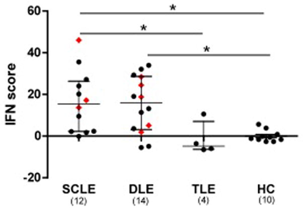
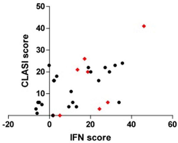
Methods: Peripheral blood was obtained from 10 healthy controls and 30 patients with CLE, including eight with concomitant SLE. Total RNA was extracted and reverse transcribed into complementary DNA. Gene expression levels were measured by real-time polymerase chain reaction. Gene expression was normalized to GAPDH, standardized to healthy controls and then summed to calculate an IFN score for each patient. Disease activity was assessed with the Cutaneous Lupus Area and Severity Index (CLASI).
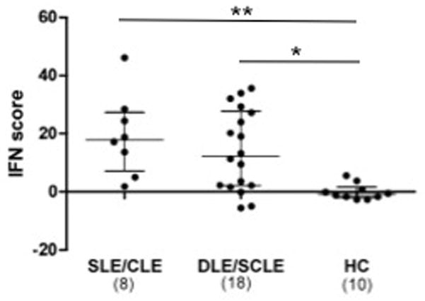
Results: Patients with subacute CLE (SCLE) and discoid lupus erythematosus (DLE) had elevated IFN scores compared with healthy controls regardless of concomitant SLE (P < 0·01 with SLE and P < 0·05 without SLE). There was no difference between patients with tumid lupus erythematosus (TLE) and healthy controls. The IFN score correlated with CLASI scores (Spearman's rho = 0·55, P = 0·0017).
Conclusions: Patients with SCLE and DLE have an IFN signature, as seen in SLE. The level of gene expression correlates with cutaneous disease activity. These findings support a shared pathogenesis between SLE and some subtypes of CLE.
© 2012 The Authors. BJD © 2012 British Association of Dermatologists.
Conflict of interest statement
There are no relevant conflicts of interest to disclose.
Figures
References
-
- Wenzel J, Zahn S, Mikus S, et al. The expression pattern of interferon-inducible proteins reflects the characteristic histological distribution of infiltrating immune cells in different cutaneous lupus erythematosus subsets. Br J Dermatol. 2007;157:752–7. - PubMed
-
- Meller S, Winterberg F, Gilliet M, et al. Ultraviolet radiation-induced injury, chemokines, and leukocyte recruitment: An amplification cycle triggering cutaneous lupus erythematosus. Arthritis Rheum. 2005;52:1504–16. - PubMed
-
- Wenzel J, Uerlich M, Haller O, et al. Enhanced type I interferon signaling and recruitment of chemokine receptor CXCR3-expressing lymphocytes into the skin following treatment with the TLR7-agonist imiquimod. J Cutan Pathol. 2005;32:257–62. - PubMed
-
- Feng X, Wu H, Grossman JM, et al. Association of increased interferon-inducible gene expression with disease activity and lupus nephritis in patients with systemic lupus erythematosus. Arthritis Rheum. 2006;54:2951–62. - PubMed
-
- Jarvinen TM, Hellquist A, Koskenmies S, et al. Tyrosine kinase 2 and interferon regulatory factor 5 polymorphisms are associated with discoid and subacute cutaneous lupus erythematosus. Exp Dermatol. 2009 - PubMed
Publication types
MeSH terms
Substances
Grants and funding
LinkOut - more resources
Full Text Sources
Other Literature Sources
Medical
Research Materials
Miscellaneous
