. 2011 Dec;119 Suppl 1(Suppl 1):199-205.
doi: 10.1111/j.1600-0722.2011.00864.x.

Matrix metalloproteinase 20 promotes a smooth enamel surface, a strong dentino-enamel junction, and a decussating enamel rod pattern

Affiliations

Matrix metalloproteinase 20 promotes a smooth enamel surface, a strong dentino-enamel junction, and a decussating enamel rod pattern

John D Bartlett et al. Eur J Oral Sci. 2011 Dec.

Abstract

Mutations of the matrix metalloproteinase 20 (MMP20, enamelysin) gene cause autosomal-recessive amelogenesis imperfecta, and Mmp20 ablated mice also have malformed dental enamel. Here we showed that Mmp20 null mouse secretory-stage ameloblasts maintain a columnar shape and are present as a single layer of cells. However, the maturation-stage ameloblasts from null mouse cover extraneous nodules of ectopic calcified material formed at the enamel surface. Remarkably, nodule formation occurs in null mouse enamel when MMP20 is normally no longer expressed. The malformed enamel in Mmp20 null teeth was loosely attached to the dentin and the entire enamel layer tended to separate from the dentin, indicative of a faulty dentino-enamel junction (DEJ). The enamel rod pattern was also altered in Mmp20 null mice. Each enamel rod is formed by a single ameloblast and is a mineralized record of the migration path of the ameloblast that formed it. The enamel rods in Mmp20 null mice were grossly malformed or absent, indicating that the ameloblasts do not migrate properly when backing away from the DEJ. Thus, MMP20 is required for ameloblast cell movement necessary to form the decussating enamel rod patterns, for the prevention of ectopic mineral formation, and to maintain a functional DEJ.


Figures

Fig. 1
Fig. 1
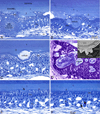
Mmp20 null mouse enamel was present in two distinct layers and had a rough surface due to the presence ectopic calcified nodules. Backscattering SEM images of the enamel surface from mandibular incisors of adult Mmp20 null mice (A, B) revealed malformed enamel. The enamel was especially malformed during the maturation stage (A; MAT) where numerous calcified nodules of irregular sizes and shapes were intermixed with cell debris (B). Semi-thin plastic demineralized histological sections (C–G) show that at the end of the secretory stage (C; SEC) the ameloblasts appear to undergo typical postsecretory transition (C, D; PST) into shorter cells typical of the maturation stage of amelogenesis. However, the enamel was thin and was composed of two layers (1, 2). Calcified nodules extend from the outermost layer nearest the ameloblasts to form a rough and uneven surface (2). Paradoxically, the innermost layer near the dentin (1) appeared relatively smooth and even (D–G). Both ameloblast and papillary layer cells were arrayed around the nodules to cover them, which distorted the spatial arrangement of the cells and, at times, made them appear folded (E, G). The nodules mineralized (F, G; min) during the maturation stage and at a somewhat faster pace than the inner two layers of enamel as observed by backscatter imaging (A, B) and the clear spaces left in the tissue following demineralization (G). The magnification bar in G applies equally to panels D–G. CEJ, cementoenamel junction; labial CT, labial connective tissue; bv, blood vessel; TP, Tomes’ Process.
Fig. 2
Fig. 2
Mmp20 null maturation stage ameloblasts form a continuous covering over the nodules that project from the enamel surface. Semi-thin plastic demineralized histological sections (A–C, E, F) and a non-demineralized section (D) show maturation stage enamel organ from mandibular incisors of adult Mmp20 null mice. The inset in panel D shows backscatter imaging of the nodules at the enamel surface. Modulating ameloblasts (Am) and associated papillary layer cells (PL) formed a continuous covering over all nodules that projected from the enamel surface in Mmp20 null mice (A–D). This caused distortions in the normal spatial arrangement of the cells perhaps giving the illusion in some cases that isolated masses of cells were swirling and/or layered (A, B) or that nodules were embedded inside the cell layers of the enamel organ (B, D). In undemineralized sections (D), nodules appeared layered with a malformed structure. Nodule-free areas were sometimes encountered in the maturation stage (E, F). The enamel in these locations often appeared thin and ameloblasts appeared somewhat distorted with small pools of protein at their apices (E, arrow). Inflammatory cell infiltration into the enamel organ was sometimes observed in sections closest to the gingival margin of the tooth (F, arrows). 1 and 2 designate the two different enamel layers. The magnification bar in B applies also to panel A; the magnification bar in F applies equally to panels C–F.
Fig. 3
Fig. 3
The dentin-enamel junction (DEJ) is weak in Mmp20 null enamel. SEM image of an incisor from an Mmp20 null mouse glued to a metal pedestal (A). Note that in the process of gluing the incisor to the pedestal a semi-conical piece of enamel fell off the dentin onto the pedestal at the base of the incisor. A semi-thin plastic demineralized histological section showing inflammatory cell infiltration into the ameloblast layer (B, red arrows). Debris appears to be accumulating at the DEJ where the enamel may already be separating from the underlying dentin (B, black arrows). d, dentin; e, enamel; g, glue; p, metal pedestal, panel B scale bar 20 µm.
Fig. 4
Fig. 4
Enamel rod patterns of mandibular incisors from wild-type and Mmp20 null mice. The wild-type enamel had crisscrossing (decussating) rows of enamel rods (A). The Mmp20 null enamel may have a poorly organized rod pattern (B), no rod pattern with a poorly organized rod layer beneath (C), or virtually no rod pattern whatsoever (D). 1 and 2 designate the two different enamel layers. Note that relative to Figures 1 and 2, the enamel from these incisors are displayed upside down. All magnification bars are 10 µm in length.

Similar articles

Cited by

References

    1. Bartlett JD, Simmer JP. Proteinases in developing dental enamel. Crit RevOral BiolMed. 1999;10:425–441. - PubMed
    1. Smith CE. Cellular and chemical events during enamel maturation. Crit RevOral BiolMed. 1998;9:128–161. - PubMed
    1. Simmer JP, Hu Y, Lertlam R, Yamakoshi Y, Hu JC. Hypomaturation enamel defects in Klk4 knockout/LacZ knockin mice. J Biol Chem. 2009;284:19110–19121. - PMC - PubMed
    1. Roycik MD, Fang X, Sang QX. A fresh prospect of extracellular matrix hydrolytic enzymes and their substrates. Curr Pharm Des. 2009;15:1295–1308. - PubMed
    1. Bartlett JD, Beniash E, Lee DH, Smith CE. Decreased mineral content in MMP-20 null mouse enamel is prominent during the maturation stage. JDentRes. 2004;83:909–913. - PubMed

Publication types

Substances