. 2012 Jan 12;73(1):92-107.
doi: 10.1016/j.neuron.2011.10.016.

Assembly and maintenance of nodes of ranvier rely on distinct sources of proteins and targeting mechanisms

Affiliations

Assembly and maintenance of nodes of ranvier rely on distinct sources of proteins and targeting mechanisms

Yanqing Zhang et al. Neuron. .

Abstract

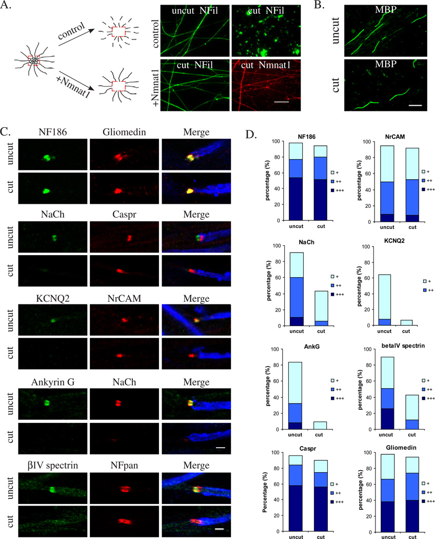
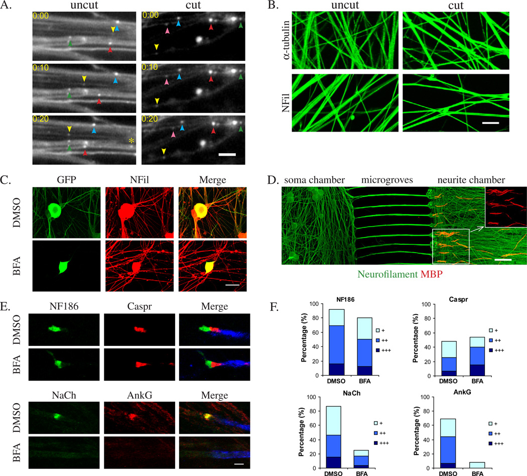
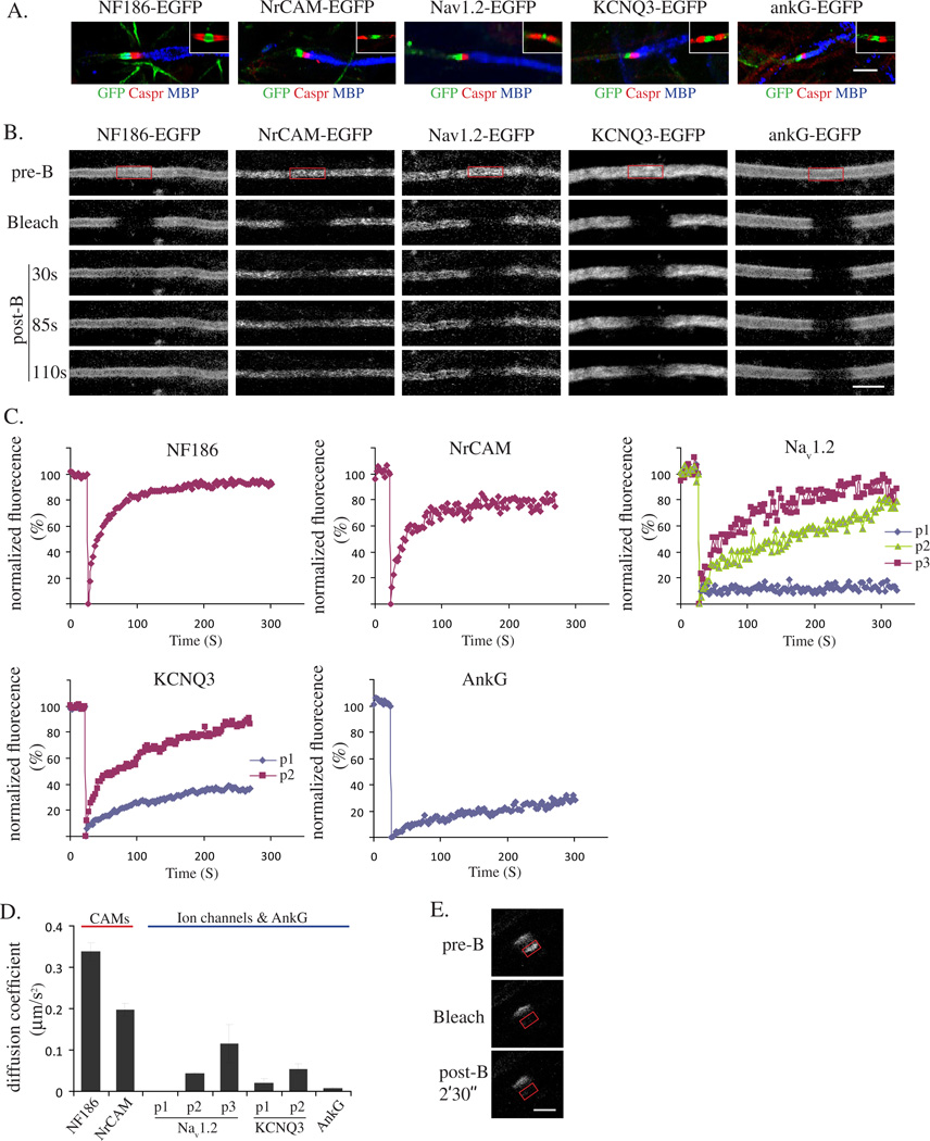
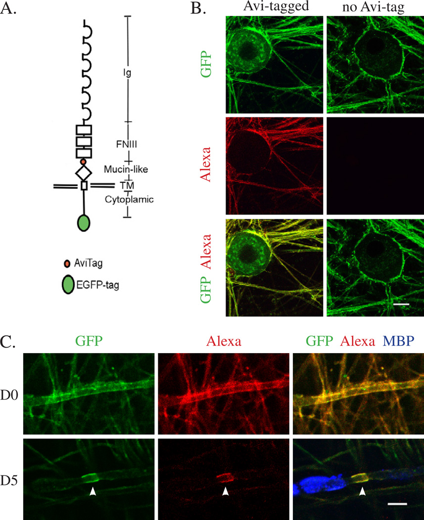
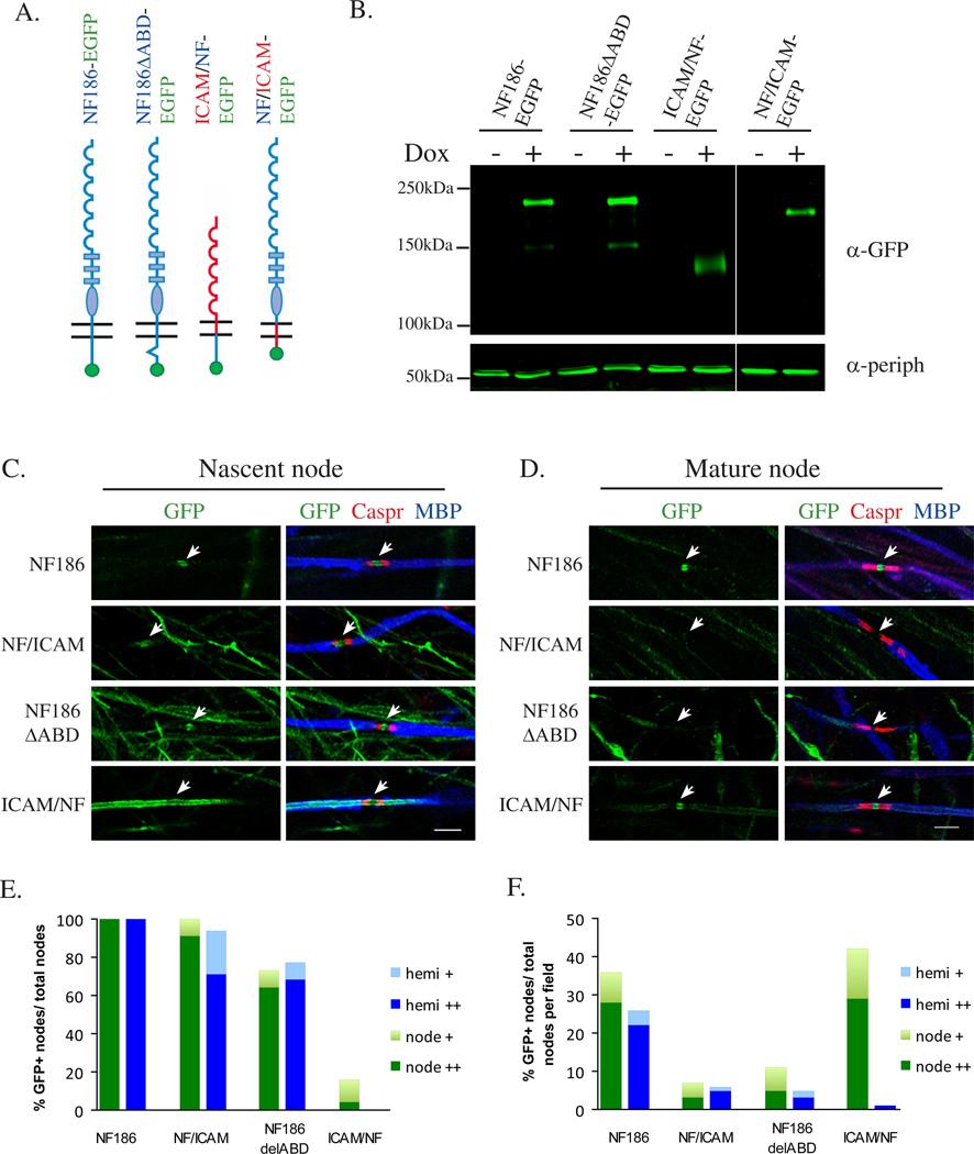
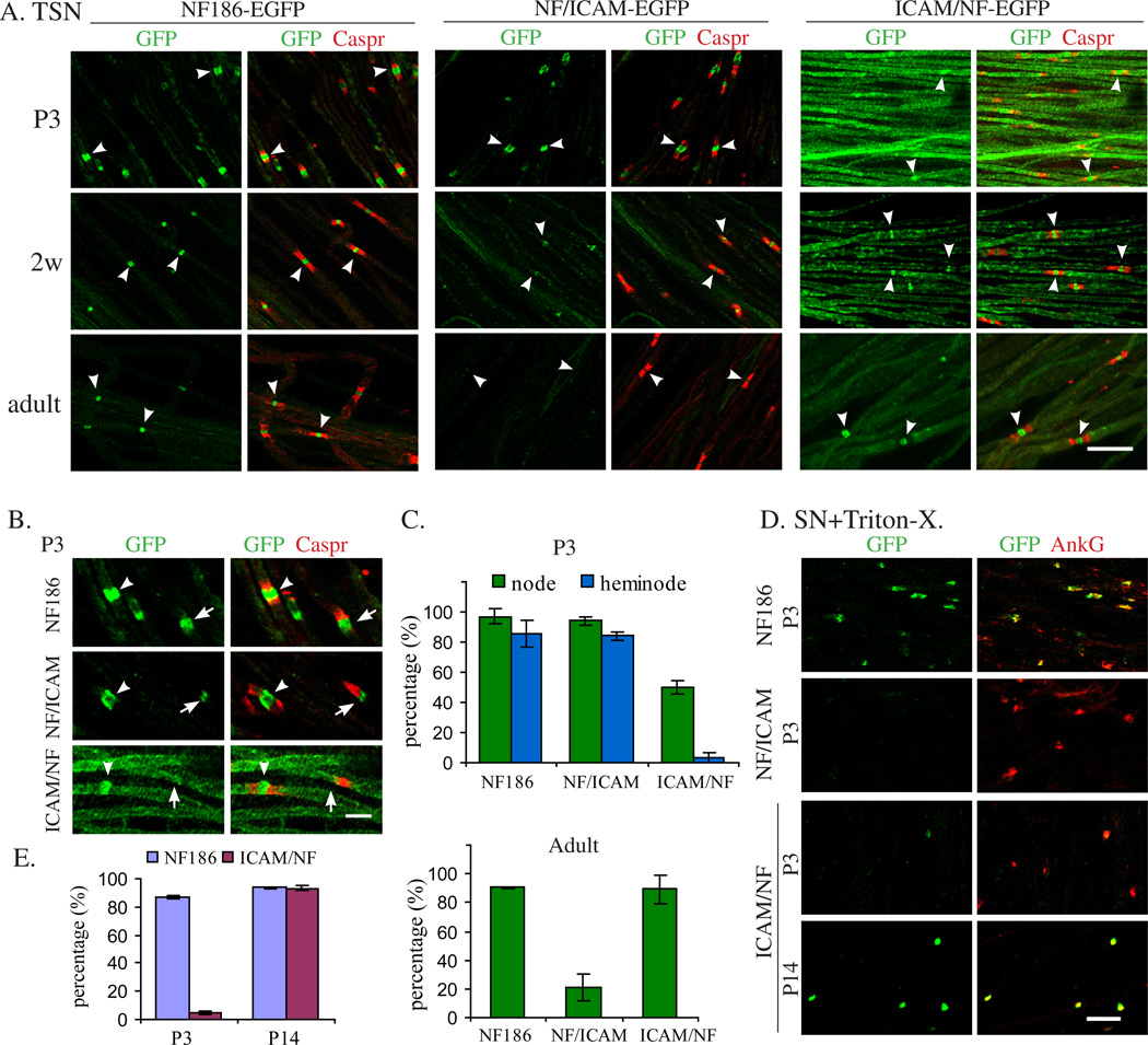
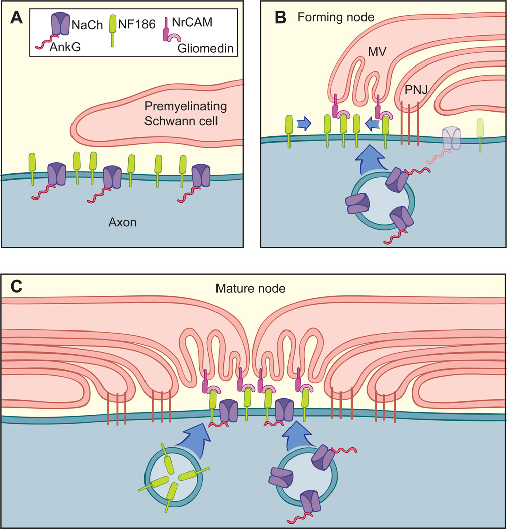
We have investigated the source(s) and targeting of components to PNS nodes of Ranvier. We show adhesion molecules are freely diffusible within the axon membrane and accumulate at forming nodes from local sources, whereas ion channels and cytoskeletal components are largely immobile and require transport to the node. We further characterize targeting of NF186, an adhesion molecule that pioneers node formation. NF186 redistributes to nascent nodes from a mobile, surface pool. Its initial accumulation and clearance from the internode require extracellular interactions, whereas targeting to mature nodes, i.e., those flanked by paranodal junctions, requires intracellular interactions. After incorporation into the node, NF186 is immobile, stable, and promotes node integrity. Thus, nodes assemble from two sources: adhesion molecules, which initiate assembly, accumulate by diffusion trapping via interactions with Schwann cells, whereas ion channels and cytoskeletal components accumulate via subsequent transport. In mature nodes, components turnover slowly and are replenished via transport.


Figures

Figure 1
Figure 1. Differential accumulation of domain components in transected Nmnat1-expressing axons
(A) Nmnat1 protects neurites from undergoing degeneration after transection. Left, schematic drawing of the experimental paradigm utilized in these experiments. Right, staining of neurites from cultures in which explants were uncut or transected 3 days beforehand from control (non-infected) or mCherry-cytNmnat1 expressing explants. Neurofilament (green) and mCherry-cytNmnat1 (red) staining are shown. Scale bar, 20 µm. (B) MBP positive myelin segments (green) formed on uncut and cut cultures protected by Nmnat1; myelination is more robust in uncut cultures. Scale bar, 50 µm. (C) Representative images showing expression of nodal and paranodal components in uncut and cut cultures; MBP segments (blue) are also shown. Axotomy was performed prior to myelination; nodal and paranodal components were examined at 7 and 11 days of myelination, respectively. Scale bar, 3 µm. (D) Quantification from a representative experiment showing the percentage of positively stained nodal and paranodal components. Positive staining was scored by intensity: very bright +++, moderately bright ++, just above background +.
Figure 2
Figure 2. Domain components accumulate by transport dependent and independent mechanisms
(A) Axonal transport rapidly fails following axotomy. Still images showing Nmnat1+ vesicles in uncut (left) and transected (right) neurites at 10 sec intervals. The positions of several vesicles are indicated by colored arrowheads; a new vesicle that entered the uncut field at 20 sec is indicated by an asterisk. Vesicles in intact neurites move at a rate of ~ 0.5 µm/sec but are stationary in the transected neurites; see also Movies S1 and S2. Scale bar, 5 µm. (B) Cytoskeleton components were not discernibly affected by axotomy. Staining of α tubulin and phospho-neurofilament in uncut and transected (6 days prior) Nmnat1+ neurites is shown. Scale bar, 8 µm. (C) Brefedin A (BFA) treatment blocks axonal transport of newly synthesized proteins. DRG neurons were infected with the doxycycline inducible lentiviral construct pSLIK-NF186-EGFP. Upon induction of NF186-EGFP, neurons were treated with either 1.0 µg/ml BFA or DMSO (vehicle control) and after 3 days, were fixed and stained for GFP (green) and neurofilament (red). NF186-EGFP was expressed in both neuronal somas and neurites in control cultures but only in the somas of BFA treated cultures. (D) Myelination in microfluidic chamber cultures. Neuronal somas and distal neurites (stained for neurofilament in green) were grown in different microfluidic culture compartments, separated by microgroves. Schwann cells were added to the neurite compartment and myelinated appropriately after 8 days in myelinating media. The inset demonstrates one field at higher magnification in order to highlight MBP-positive segments; for clarity only the red channel is shown. Scale bar, 100 µm. (E) BFA treatment blocked nodal accumulation of sodium channels and ankyrin G but not NF186 and Caspr. BFA was added to the compartment containing neuronal somas prior to initiating myelination in the neurite compartment. Representative images showing expression of nodal and paranodal components in control (DMSO) and BFA treated cultures (7 days of myelination, the last 5 with DMSO or BFA); MBP segments (blue) are also shown. Scale bar, 3 µm. (F) Quantification from a representative experiment showing the percentage of positively stained nodal and paranodal components. Positive staining was score by intensity: very bright +++, moderately bright ++, just above background +.
Figure 3
Figure 3. Differential lateral mobility of nodal components
(A) EGFP-tagged constructs of nodal components are appropriately targeted to nodes and heminodes. Constructs were nucleofected into neurons, which were then cocultured with Schwann cells under myelinating conditions for 2 weeks. These constructs, identified by staining for GFP, are all targeted appropriately to heminodes and nodes (inset); paranodes were identified by staining for Caspr (red) and myelin segments with MBP (blue). Scale bar, 6 µm. (B) Representative images showing results of photobleaching and subsequent recovery of GFP fluorescence. The GFP-tagged constructs shown in panel A were expressed in DRG neurons. Cultures were treated with nocodazole and single, labeled neurites were photobleached (site delineated by the red box); the extent of the recovery of fluorescence was observed at subsequent time intervals. (C) Representative examples of FRAP analysis for NF186, NrCAM, Nav1.2, KCNQ3 and ankyrin G. Note, NF186 and NrCAM are uniformly mobile; mobilities of Nav1.2 and KCNQ3 were significantly reduced and more variable with three broad patterns (p1 to 3) detected for Nav1.2 and two (p1 and 2) detected for KCNQ3; ankyrin G is essentially immobile. (D) Summary of the diffusion coefficients measured for each component. (E) NF186 incorporated into the node is effectively immobile. One half of a node in which NF186-EGFP is concentrated (which appears as two linear signals due edge effects) was photobleached (demarcated by the red box). No recovery was detected even 2 ½ minutes after photobleaching. Scale bar, 2 µm.
Figure 4
Figure 4. Neurofascin on the axon surface redistributes to forming nodes of Ranvier
(A) Schematic drawing of the NF186 construct showing the locations of the AviTag and the EGFP-tag. (B) The AviTag construct was detectably expressed in DRG neurons (GFP), surface biotinylated by addition of BirA ligase, and visualized with streptavidin conjugated to AlexaFluor 568 (Alexa). NF186-EGFP without an AviTag was not biotinylated. Scale bar, 15 µm. (C) DRG neurons expressing the AviTagged NF186-EGFP were biotinylated, then cocultured with Schwann cells under myelinating condition. The total (GFP) and biotinylated (identified by Alexa staining) populations of AviTagged NF186-EGFP were readily detected all along the neurite immediately after biotinylation (D0 panel). 5 days later this surface pool was confined to newly formed nodes (D5 panel); unmyelinated fibers at D5 express much lower levels of the biotinylated construct. Scale bar, 4 µm.
Figure 5
Figure 5. NF186 incorporated into nodes is stable and maintains sodium channel expression
(A) Representative images showing the effect of knocking down NF186 prior to (left panels) vs. after (right panels) nodes of Ranvier have formed. Cultures were infected with lentiviral constructs encoding shRNA to NF186 or control (scrambled, Scr) sequences and stained for NF186 (red) and Caspr (blue); nerve fibers infected with the lentiviral constructs are GFP+ (green). Left panels: NF186 was not detected at nodes when neurons were treated with shRNA 3 wks prior to the onset of myelination. Right panels: NF186 continued to be robustly expressed at nodes (arrowheads) when neurons were treated with shRNA 6 weeks after myelination had commenced and continuing for one month more; expression at heminodes (arrow) was slightly reduced. Scale bar, 10 µm. (B) The relative intensities of NF186 at heminodes and nodes of neurons treated with shRNA to NF186 vs. scrambled (control) sequences were determined following shRNA treatment; knockdown of NF186 by the shRNA construct was initiated 6 weeks after myelination and continued for the times shown. (C) The relative intensities of NaCh at heminodes and nodes of neurons treated with shRNA to NF186 vs. scrambled (control) sequences are shown at various times following shRNA treatment of the cultures in B. Results shown in B and C are averaged from two sets of experiments; error bars correspond to the average deviation.
Figure 6
Figure 6. NF186 is targeted to forming and mature nodes by different mechanisms
(A) Schematic drawing of wild type, mutant, and chimeric NF186 constructs used in the experiment; NF186 domains are shown in blue, ICAM domains are in red. (B) Inducible expression of NF constructs in DRG neurons is doxycycline (1 µg/ml) dependent; peripherin is used as a loading control. (C) Representative images of the targeting of different constructs to nascent node (indicated by white arrows). Doxycycline was added to cultures prior to switching to myelinating media; these were maintained under myelinating conditions for an additional 7– 10 days. Scale bar, 6 µm. (D) Representative images of the targeting of different constructs to mature nodes (indicated by white arrows). Doxycycline was added to cocultures that had already myelinated for 6 weeks; these were then maintained for an additional 1 to 2 weeks. (E) Quantification of the targeting of constructs to nascent nodes is presented as a percentage of the total nodes on GFP-positive fibers. (F) Quantification of the targeting of constructs to mature nodes is presented as the ratio of positively nucleated nodes vs. total nodes in the scanned fields.
Figure 7
Figure 7. Targeting of NF186 constructs in peripheral nerves of transgenic mice
(A) Teased sciatic nerves (TSN) from wt NF186 (EGFP), NF/ICAM and ICAM/NF transgenic mouse lines were stained for GFP and Caspr (red) at the different ages shown. Scale bar, 20 µm. (B) Higher power images showing targeting to nodes (arrowheads) vs. heminodes (arrows) of transgenic constructs at P3. Scale bar, 5 µm. (C) Quantification of GFP positive nodes and heminodes in P3 and GFP positive nodes in adult TSN, shown as percentage of total nodes or heminodes; standard deviations are shown. NF/ICAM-positive nodes in adult sciatic nerves only faintly expressed this transgene in most cases. (D) Sciatic nerves from P3 NF186 and NF/ICAM transgenic mice and from P3 and P14 ICAM/NF transgenic mouse were extracted with 0.5% Triton X-100 then fixed and stained for GFP and ankyrin G. Scale bar, 10 µm. (E) Quantification of results for P3 and P14 comparing wt NF186 and ICAM/NF from Triton extracted sciatic nerves; standard deviations are shown.
Figure 8
Figure 8. Multiple mechanisms contribute to node assembly and maintenance
(A) Prior to myelination, components of the node and other domains (not shown) are diffusely expressed along the axon; NF186 is not associated with ankyrin G. (B) Node assembly: NF186 on the axon surface redistributes via diffusion to the node where it is “trapped” by interactions with the gliomedin/NrCAM complex on Schwann cell microvilli (MV). Sodium channels, which are shown complexed in some cases to ankyrin G in transport vesicles, are delivered for exocytosis; whether these components all traffic by fast transport and are delivered together or separately is not yet known. Nodal components along the internode are also downregulated (transparent components); early paranodal junctions (PNJ) are also illustrated. (C) In mature nodes, all node components are shown being delivered by transport of carrier vesicles, replenishing components that slowly turnover. At the node, NF186 is linked to sodium channels via ankyrin G. The flanking paranodal junctions provide a lateral diffusion barrier and may direct targeting to the node. While NF186 is illustrated as being in separate transport vesicles from those carrying sodium channels and ankyrin G, the precise composition of transport vesicles remains to be established, including whether ankyrin is transported separately by slow transport. NrCAM and other components of the node are not shown to simplify the figure.

Comment in

Similar articles

Cited by

References

    1. Aigner L, Arber S, Kapfhammer JP, Laux T, Schneider C, Botteri F, Brenner HR, Caroni P. Overexpression of the neural growth-associated protein GAP-43 induces nerve sprouting in the adult nervous system of transgenic mice. Cell. 1995;83:269–278. - PubMed
    1. Alix JJ, Dolphin AC, Fern R. Vesicular apparatus, including functional calcium channels, are present in developing rodent optic nerve axons and are required for normal node of Ranvier formation. J Physiol. 2008;586:4069–4089. - PMC - PubMed
    1. Anderson E, Maday S, Sfakianos J, Hull M, Winckler B, Sheff D, Folsch H, Mellman I. Transcytosis of NgCAM in epithelial cells reflects differential signal recognition on the endocytic and secretory pathways. J Cell Biol. 2005;170:595–605. - PMC - PubMed
    1. Armstrong R, Toews AD, Morell P. Axonal transport through nodes of Ranvier. Brain Res. 1987;412:196–199. - PubMed
    1. Beckett D, Kovaleva E, Schatz PJ. A minimal peptide substrate in biotin holoenzyme synthetase-catalyzed biotinylation. Protein Sci. 1999;8:921–929. - PMC - PubMed

Publication types

MeSH terms