. 2012 Jan 13:12:15.
doi: 10.1186/1471-2407-12-15.

Multicellular tumor spheroid model to evaluate spatio-temporal dynamics effect of chemotherapeutics: application to the gemcitabine/CHK1 inhibitor combination in pancreatic cancer

Affiliations

Multicellular tumor spheroid model to evaluate spatio-temporal dynamics effect of chemotherapeutics: application to the gemcitabine/CHK1 inhibitor combination in pancreatic cancer

Isabelle Dufau et al. BMC Cancer. .

Abstract

Background: The multicellular tumor spheroid (MCTS) is an in vitro model associating malignant-cell microenvironment and 3D organization as currently observed in avascular tumors.

Methods: In order to evaluate the relevance of this model for pre-clinical studies of drug combinations, we analyzed the effect of gemcitabine alone and in combination with the CHIR-124 CHK1 inhibitor in a Capan-2 pancreatic cell MCTS model.

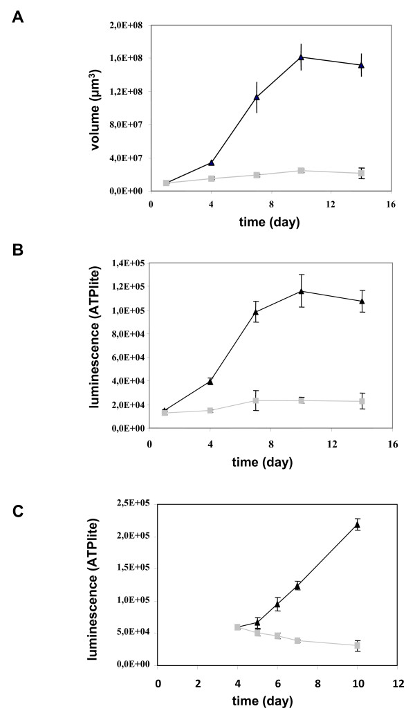
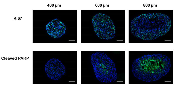
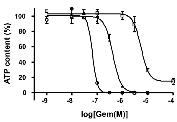
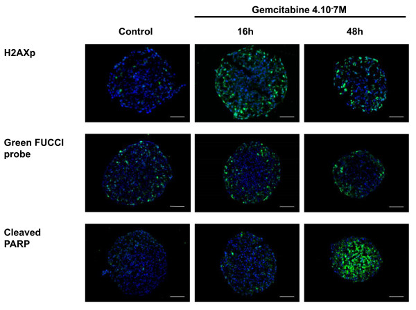
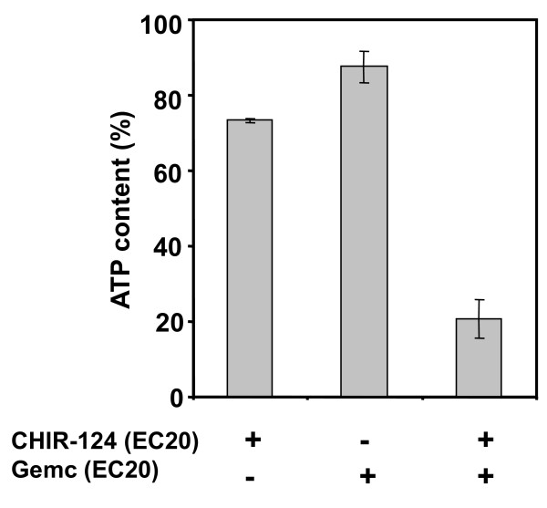
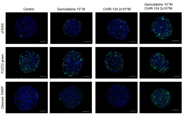
Results: Compared to monolayer cultures, Capan-2 MCTS exhibited resistance to gemcitabine cytotoxic effect. This resistance was amplified in EGF-deprived quiescent spheroid suggesting that quiescent cells are playing a role in gemcitabine multicellular resistance. After a prolonged incubation with gemcitabine, DNA damages and massive apoptosis were observed throughout the spheroid while cell cycle arrest was restricted to the outer cell layer, indicating that gemcitabine-induced apoptosis is directly correlated to DNA damages. The combination of gemcitabine and CHIR-124 in this MCTS model, enhanced the sensitivity to the gemcitabine antiproliferative effect in correlation with an increase in DNA damage and apoptosis.

Conclusions: These results demonstrate that our pancreatic MCTS model, suitable for both screening and imaging analysis, is a valuable advanced tool for evaluating the spatio-temporal effect of drugs and drug combinations in a chemoresistant and microenvironment-depending tumor model.


Figures

Figure 1
Figure 1
Growth of Capan-2 spheroids. The Capan-2 spheroids were grown in the presence of 10% serum (gray squares) or defined medium added with EGF and B27 (black triangles) without any medium change (a and b). (a) Spheroid size. The diameter of each spheroid was measured by microscopic observation with an ocular micrometer and sphere volume was calculated. (b) Cell viability. Spheroid viability was quantified by ATP monitoring with the luminescent ATPlite assay. (c) Induction of a quiescent state on Capan-2 spheroid. Capan-2 spheroid culture was initiated in presence of 10% serum and EGF and four days after culture initiation, EGF was (gray squares) or not (black triangles) removed from the culture medium. Capan-2 spheroid viability was followed during a 6 days additional period with the luminescent ATPlite assay. Each point is the mean ± SD of 6 spheroids.
Figure 2
Figure 2
Distribution of proliferative and apoptotic cells in Capan-2 spheroids of different sizes. Ki-67 and cleaved form of PARP were analyzed by immunodetection on 5 μm frozen sections in Capan-2 spheroids of different size. 400, 600 and 800 μm correspond respectively to spheroid at days 4, 7, and 12 after culture initiation in defined medium supplemented with EGF and B27. The scale bar corresponds to 100 μm. Results shown are representative of the examination of 3 sections from 5 spheroids. Each experiment has been repeated 3 times.
Figure 3
Figure 3
Gemcitabine effect on cell viability after 72 h treatment on Capan-2 cells cultured as monolayer (open circle), spheroid in defined medium supplemented with EGF and B27 (black triangles), or as quiescent Capan-2 spheroid (gray squares). Each point is the mean ± SD of 3 replicates. Dose-response experiments were fitted with GraphPad Prism software.
Figure 4
Figure 4
Spatio-temporal response of Capan-2 spheroid to gemcitabine. Analysis of gemcitabine response was done on 5 μm frozen sections of Capan-2 spheroids treated 16 h and 48 h with 4.10-7 M gemcitabine. DNA damage was revealed by immunodetection of phosphorylated of γH2AX, S phase checkpoint was monitored on Capan-2 spheroid expressing the geminin-mAG FUCCi green probe and apoptosis was analyzed by immunodetection of cleaved form of PARP. Images were collected using a X10 objective. The scale bar corresponds to 100 μm. Results shown are representative of the examination of 3 sections from 5 spheroids. Each experiment has been repeated 3 times.
Figure 5
Figure 5
Gemcitabine and CHIR-124 produce a synergistic cytotoxic effect on Capan-2 spheroid. Spheroids were treated for 72 h after 4 day initial culture with gemcitabine, CHIR-124 or a combination of the 2 compounds at concentration corresponding to their respective EC20 values. Spheroid viability was quantified at day 7 by ATP monitoring with the luminescent ATPlite assay. ATP content percentage was calculated with regard to non-treated spheroid and showed cell growth inhibition and/or toxicity.
Figure 6
Figure 6
Spatio-temporal response of Capan-2 spheroid to gemcitabine and Chk1 inhibitor. Analysis of gemcitabine response in presence and absence of CHK1 inhibitor was done on 5 μm frozen sections of Capan-2 spheroids treated 16 h with 10-7 M gemcitabine alone, 2 10-8 M CHIR-124 alone or the combination of gemcitabine and CHIR-124 at these same doses. DNA damage was revealed by immunodetection of phosphorylated of γH2AX, S phase checkpoint was monitored on Capan-2 spheroid expressing the geminin-mAG Fucci green probe and apoptosis was analyzed by immunodetection of cleaved form of PARP. Images were collected using a X10 objective. The scale bar corresponds to 100 μm. Results shown are representative of the examination of 3 sections from 5 spheroids. Each experiment has been repeated 3 times.

References

    1. Azorsa DO, Gonzales IM, Basu GD, Choudhary A, Arora S, Bisanz KM, Kiefer JA, Henderson MC, Trent JM, Von Hoff DD, Mousses S. Synthetic lethal RNAi screening identifies sensitizing targets for gemcitabine therapy in pancreatic cancer. J Transl Med. 2009;7:43. doi: 10.1186/1479-5876-7-43. - DOI - PMC - PubMed
    1. Ma CX, Janetka JW, Piwnica-Worms H. Death by releasing the breaks. CHK1 inhibitors as cancer therapeutics. Trends Mol Med. 2011;17:88–96. doi: 10.1016/j.molmed.2010.10.009. - DOI - PMC - PubMed
    1. Matthews DJ, Yakes FM, Chen J, Tadano M, Bornheim L, Clary DO, Tai A, Wagner JM, Miller N, Kim YD, Robertson S, Murray L, Karnitz LM. Pharmacological abrogation of S-phase checkpoint enhances the anti-tumor activity of gemcitabine in vivo. Cell Cycle. 2007;6:104–110. doi: 10.4161/cc.6.1.3699. - DOI - PubMed
    1. Parsels LA, Morgan MA, Tanska DM, Parsels JD, Palmer BD, Booth RJ, Denny WA, Canman CE, Kraker AJ, Lawrence TS, Maybaum J. Gemcitabine sensitization by checkpoint kinase 1 inhibition correlates with inhibition of a Rad51 DNA damage response in pancreatic cancer cells. Mol Cancer Ther. 2009;8:45–54. doi: 10.1158/1535-7163.MCT-08-0662. - DOI - PMC - PubMed
    1. Morgan MA, Parsels LA, Zhao L, Parsels JD, Davis MA, Hassan MC, Arumugarajah S, Hylander-Gans L, Morosini D, Simeone DM, Canman CE, Normolle DP, Zabludoff SD, Maybaum J, Lawrence TS. Mechanism of radiosensitization by the Chk1/2 inhibitor AZD7762 involves abrogation of the G2 checkpoint and inhibition of homologous recombinational DNA repair. Cancer Res. 2010;70:4972–4981. doi: 10.1158/0008-5472.CAN-09-3573. - DOI - PMC - PubMed

Publication types

MeSH terms