. 2012 Jan 13:2:2.
doi: 10.1186/2045-3701-2-2.

Human papillomavirus 18 E6 inhibits phosphorylation of p53 expressed in HeLa cells

Affiliations

Human papillomavirus 18 E6 inhibits phosphorylation of p53 expressed in HeLa cells

Amrendra K Ajay et al. Cell Biosci. .

Abstract

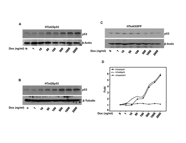
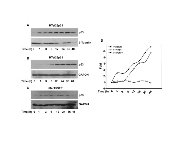
Background: In HPV infected cells p53 function is abrogated by E6 and even ectopically expressed p53 is unable to perform tumor suppressor functions. In addition to facilitating its degradation, E6 may also inhibit p53 transactivity, though the mechanisms are still poorly understood. It has been reported that inhibition of p300, an acetyltransferase responsible for p53 acetylation is inactivated by E6. Activation of overexpressed p53 to cause cell growth inhibition is facilitated by its phosphorylation. Previously, we reported that non-genotoxically overexpressed p53 in HeLa cells needs to be phosphorylated to perform its cell growth inhibitory functions. Since over expressed p53 by itself was not activated, we hypothesized an inhibitory role for E6.

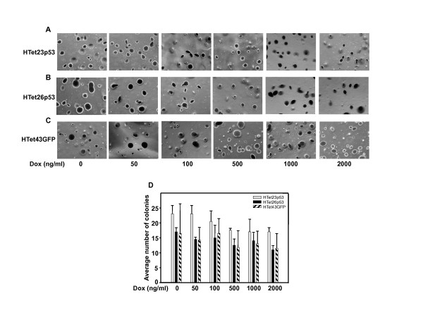
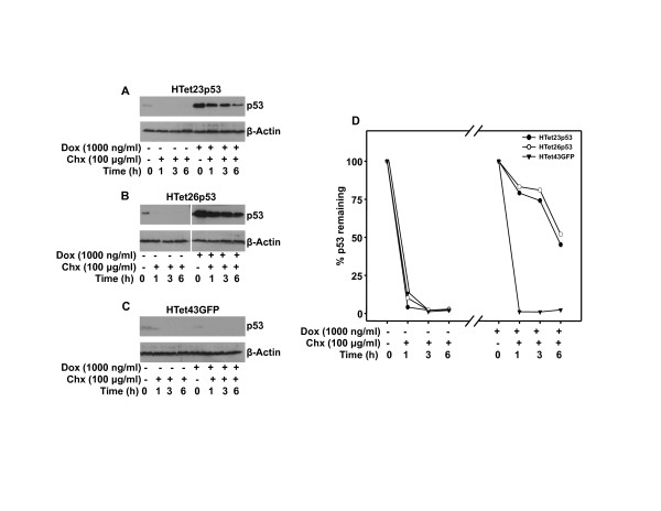
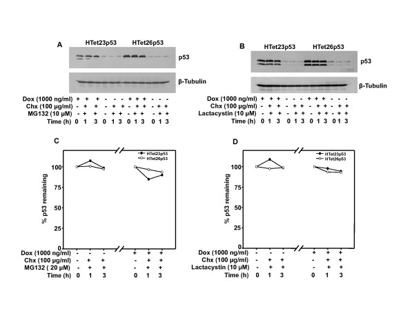
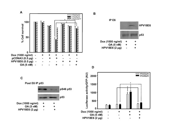
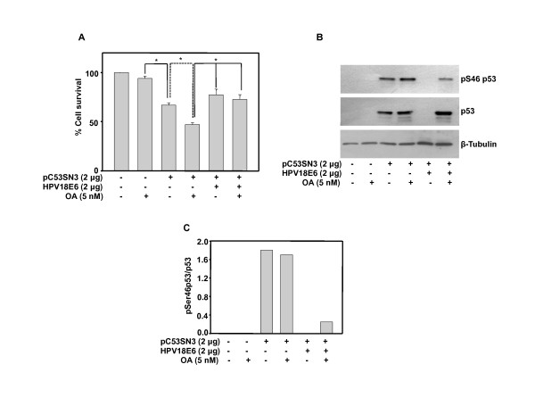
Results: Majority of reports proposes E6 mediated degradation of p53 as a possible reason for its inactivation. However, results presented here for the first time demonstrate that overexpressed p53 is not directly associated with E6 and therefore free, yet it is not functionally active in HPV positive cells. Also, the stability of overexpressed p53 does not seem to be an issue because inhibition of proteasomal degradation did not increase the half-life of overexpressed p53, which is more than endogenous p53. However, inhibition of proteasomal degradation prevents the degradation of endogenous p53. These findings suggest that overexpressed p53 and endogenous p53 are differentially subjected to proteasomal degradation and the reasons for this discrepancy remain unclear. Our studies demonstrate that p53 over expression has no effect on anchorage independent cell-growth and E6 nullifies its cell growth inhibitory effect. E6 overexpression abrogates OA induced p53 occupancy on the p21 promoter and cell death as well. E6 did not decrease p53 protein but phospho-p53 level was significantly reduced.

Conclusion: We report for the first time that E6 de-activates p53 by inhibiting its phosphorylation. This prevents p53 binding to p21 promoter and thereby restraining its cell-growth inhibitory functions. Our study provides new evidence indicating that viral protein E6 inhibits p53 transactivity by mechanism independent of degradation pathway.


Figures

Figure 1
Figure 1
p53 expression is regulated in a dose dependent manner by Dox. (A and B) Cells treated with Dox for 48 h were harvested and western blot for p53 was performed. (C) No induction of p53 in response to Dox was observed in HTet43GFP cells. (D) Fold induction of p53 was calculated by densitometric analysis of p53 western blot in HTet23p53, HTet26p53 and HTet43GFP cells taking control = 1with normalization to β-Actin or β -Tubulin.
Figure 2
Figure 2
p53 expression is regulated in a time dependent manner. (A, B and C) HTet23p53, HTet26p53 and HTet43GFP cells were treated with 1000 ng/ml Dox for indicated time points and western blots were performed for p53. (D) Fold induction was calculated by densitometric analysis of p53 in HTet23p53, HTet26p53 and HTet43GFP cells taking 0 h = 1 with normalization to β -Tubulin or GAPDH.
Figure 3
Figure 3
p53 over expression did not reduce colony number. Five thousand cells were plated in low-melting agarose containing complete medium and allowed to grow for 30 days. (A, B and C) Cells were stained with crystal violet and photographs were taken under microscope for HTet23p53, HTet26p53 and HTet43GFP plates. (D) Colonies were counted and average number of colonies was plotted vs Dox concentration. Bar represents average of colonies from two representative fields (± SE).
Figure 4
Figure 4
Overexpressed p53 is stable. (A and B) p53 was overexpressed with 1000 ng/ml of Dox for 48 h or not overexpressed and western blotswere performed after indicated time of Chx treatment in HTet23p53 and HTet26p53 cells. (C) Western blot for p53 was performed with or without addition of 1000 ng/ml Dox for 48 h followed by Chx treatment for indicated time points in HTet43GFP cells.(D) Graphical representation of percentage of p53 protein remaining after indicated time points in HTet23p53 and HTet26p53 cells by densitometric analysis following normalization with β-Actin. Protein percentage for 0 h Chx was taken as 100.
Figure 5
Figure 5
Overexpressed p53 is not stabilized by inhibition of proteasomal degradation. Cells were treated with proteasomal inhibitors (MG132 or Lactacystin) 1 h prior to Dox addition and incubated for 48 h. Thereafter, cells were treated with Chx for indicated time points and western blots were performed. (A) MG132 treatment in HTet23p53 and HTet26p53 cells and p53 protein expression. (B) Lactacystin treatment in HTet23p53 and HTet26p53 cells and p53 protein expression.
Figure 6
Figure 6
Overexpressed p53 is functionally impaired by HPV E6. (A) HTet23p53, HTet26p53 or HTet43GFP cells were transfected with vector or HPV18 E6 plasmid and 18 h post transfection cells were treated with OA 1 h prior to Dox addition. MTT assay was performed after 48 h. Bar represents variations among the wells of an experiment done twice in triplicate. * Indicates P < 0.01 (B) HTet26p53 cells were transfected with HPV18 E6 plasmid and treated as mentioned in A and immunoprecipitation was performed first with E6 antibody (first IP) and secondly by p53 antibody. p53 or E6 was detected in immunoprecipitated complex. (C) p53 post-IP (second IP) following E6 IP was performed and p53 or phospho-p53detection by western blotting was performed. (D) Cells were transfected with p21luciferase construct with or without HPV18 E6 plasmid and treated as mentioned in A. Luciferase assay was performed and luciferase/GFP reading was plotted. Bar represents results from an experiment done in triplicate. (± SE). * Indicates P < 0.05.
Figure 7
Figure 7
Overexpressed p53 is active and is made non-functional by HPV 18 E6 in H1299, a p53 and E6 null cell line. (A) H1299 cells were transfected with pC53-SN3 and/or HPV18 E6 plasmids and 18 h post transfection cells were treated with OA. MTT assay was performed after 48 h. Bar represents variations among the wells of an experiment done twice in triplicate. * Indicates P < 0.01 (B) H1299 cells were transfected with pC53-SN3 and/or HPV18E6 plasmids and 18 h post transfection cells were treated with OA for 48 h and western blot was performed for pSer46p53 and p53.(C) Densitometric analysis was performed by normalization with p53 and ratio of pSer53 and p53 was plotted.

References

    1. Walboomers JM, Jacobs MV, Manos MM, Bosch FX, Kummer JA, Shah KV, Snijders PJ, Peto J, Meijer CJ, Munoz N. Human papillomavirus is a necessary cause of invasive cervical cancer worldwide. J Pathol. 1999;189:12–19. doi: 10.1002/(SICI)1096-9896(199909)189:1<12::AID-PATH431>3.0.CO;2-F. - DOI - PubMed
    1. Franceschi S, Munoz N, Bosch XF, Snijders PJ, Walboomers JM. Human papillomavirus and cancers of the upper aerodigestive tract: a review of epidemiological and experimental evidence. Cancer Epidemiol Biomarkers Prev. 1996;5:567–575. - PubMed
    1. Brandwein M, Zeitlin J, Nuovo GJ, MacConnell P, Bodian C, Urken M, Biller H. HPV detection using "hot start" polymerase chain reaction in patients with oral cancer: a clinicopathological study of 64 patients. Mod Pathol. 1994;7:720–727. - PubMed
    1. Fouret P, Monceaux G, Temam S, Lacourreye L, St Guily JL. Human papillomavirus in head and neck squamous cell carcinomas in nonsmokers. Arch Otolaryngol Head Neck Surg. 1997;123:513–516. doi: 10.1001/archotol.1997.01900050063008. - DOI - PubMed
    1. Gillison ML, Koch WM, Shah KV. Human papillomavirus in head and neck squamous cell carcinoma: are some head and neck cancers a sexually transmitted disease? Curr Opin Oncol. 1999;11:191–199. doi: 10.1097/00001622-199905000-00010. - DOI - PubMed