. 2012 Jan 13:12:3.
doi: 10.1186/1472-6882-12-3.

Screening of anti-dengue activity in methanolic extracts of medicinal plants

Affiliations

Screening of anti-dengue activity in methanolic extracts of medicinal plants

Leon I C Tang et al. BMC Complement Altern Med. .

Abstract

Background: Dengue fever regardless of its serotypes has been the most prevalent arthropod-borne viral diseases among the world population. The development of a dengue vaccine is complicated by the antibody-dependent enhancement effect. Thus, the development of a plant-based antiviral preparation promises a more potential alternative in combating dengue disease.

Methods: Present studies investigated the antiviral effects of standardised methanolic extracts of Andrographis paniculata, Citrus limon, Cymbopogon citratus, Momordica charantia, Ocimum sanctum and Pelargonium citrosum on dengue virus serotype 1 (DENV-1).

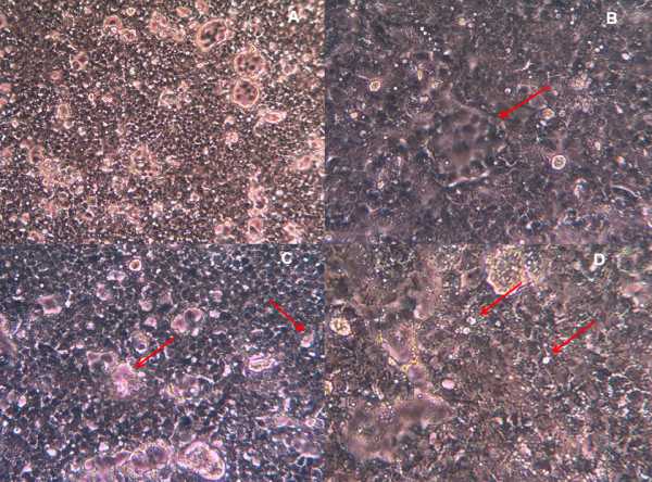
Results: O. sanctum contained 88.6% of total flavonoids content, an amount that was the highest among all the six plants tested while the least was detected in M. charantia. In this study, the maximum non-toxic dose (MNTD) of the six medicinal plants was determined by testing the methanolic extracts against Vero E6 cells in vitro. Studies also determined that the MNTD of methanolic extract was in the decreasing order of M. charantia >C. limon >P. citrosum, O. sanctum >A. paniculata >C. citratus. Antiviral assay based on cytopathic effects (CPE) denoted by degree of inhibition upon treating DENV1-infected Vero E6 cells with MNTD of six medicinal plants showed that A. paniculata has the most antiviral inhibitory effects followed by M. charantia. These results were further verified with an in vitro inhibition assay using MTT, in which 113.0% and 98.0% of cell viability were recorded as opposed to 44.6% in DENV-1 infected cells. Although methanolic extracts of O. sanctum and C. citratus showed slight inhibition effect based on CPE, a significant inhibition was not reflected in MTT assay. Methanolic extracts of C. limon and P. citrosum did not prevent cytopathic effects or cell death from DENV-1.

Conclusions: The methanol extracts of A. paniculata and M. charantia possess the ability of inhibiting the activity of DENV-1 in in vitro assays. Both of these plants are worth to be further investigated and might be advantageous as an alternative for dengue treatment.


Figures

Figure 1
Figure 1
The percentage of toxicity of methanolic extracts of six plants on Vero E6 cells after incubated in vitro for 96 hours. (A) Momordica charantia (B) Cymbopogon citratus (C) Andrographis paniculata (D) Citrus limon (E) Pelargonium citrosum (F) Ocimum sanctum. The data shown are means ± S.D. of two independent experiments performed in triplicates.
Figure 2
Figure 2
A monolayer sheet of uninfected Vero E6 cells. The normal cells were polygonal with well defined, black nuclei in the centre. The cytoplasm was shaded grey with the cell membranes were clearly demarcated with white lines surrounding the cells. The cells were viewed under inverted microscope at 200× magnification.
Figure 3
Figure 3
Various cytopathic effects observed in DENV-1-infected Vero E6 cells after 7 days post infection. (A) The monolayer sheet of cells at 100× magnification appeared to be congested and haphazard. (B) A typical syncythial giant cell formation or syncytia resulting from the fusion of a few cells infected with DENV-1 viewed at 200× magnification. The morphological changes appeared to be group of cells without a clear white demarcation of cell membrane. The cells located at the perimeter of the syncytia were observed clearly. (C) Areas of cell death or lysis are pointed out by the arrows. These small areas were clear of any cells and reflected the colour of the pink medium. (D) A higher magnification image (400×) showing white dots called blebs.
Figure 4
Figure 4
Morphological changes of DENV-1-infected Vero E6 cells treated with methanolic extracts of six medicinal plants at 7 days post infection. All the cells were viewed under inverted microscope at 200× magnification. (A) A. paniculata with75% of inhibition. (B) M. charantia with 50% of inhibition. (C) O. sanctum with less than 50% inhibition. (D) C. citratus with less than 50% inhibition (E) C. limon with no inhibition. Only cell fragments and debris were seen (F) P. citrosum with no inhibition. Granulation and cell densing were observed. The CPE for each treatment was compared with those of negative and positive controls shown in Figures 2 and 3, respectively.

References

    1. Halstead SB. Dengue virus - Mosquito interactions. Annu Rev Entomol. 2008;53:273–291. doi: 10.1146/annurev.ento.53.103106.093326. - DOI - PubMed
    1. Guzman MG, Kouri G. Dengue diagnosis, advances and challenges. Int J Infect Dis. 2004;8:69–80. doi: 10.1016/j.ijid.2003.03.003. - DOI - PubMed
    1. Gibbons RV. Dengue conundrums. Int J Antimicrob Agents. 2010;365:S36–S39. - PMC - PubMed
    1. Gubler DJ. Dengue and dengue hemorrhagic fever. Clin Microbiol Rev. 1998;11:480–96. - PMC - PubMed
    1. Murrell S, Wu SC, Butler M. Review of dengue virus and the development of a vaccine. Biotechnol Adv. 2011;29:239–247. doi: 10.1016/j.biotechadv.2010.11.008. - DOI - PubMed

Publication types

MeSH terms