Review
. 2012 Apr;133(4):195-202.
doi: 10.1016/j.mad.2011.11.009. Epub 2012 Jan 8.

Double strand binding-single strand incision mechanism for human flap endonuclease: implications for the superfamily

Affiliations
Review

Double strand binding-single strand incision mechanism for human flap endonuclease: implications for the superfamily

Susan E Tsutakawa et al. Mech Ageing Dev. 2012 Apr.

Abstract

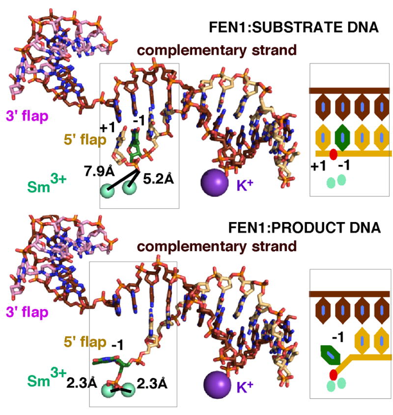
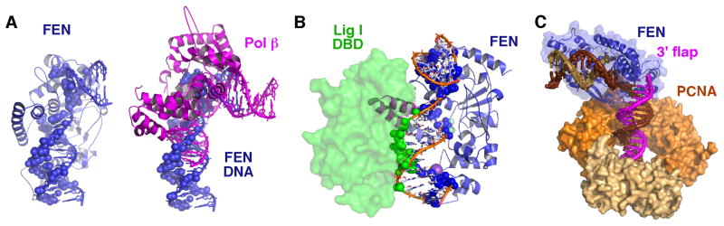
Detailed structural, mutational, and biochemical analyses of human FEN1/DNA complexes have revealed the mechanism for recognition of 5' flaps formed during lagging strand replication and DNA repair. FEN1 processes 5' flaps through a previously unknown, but structurally elegant double-stranded (ds) recognition/single stranded (ss) incision mechanism that both selects for 5' flaps and selects against ss DNA or RNA, intact dsDNA, and 3' flaps. Two major DNA binding interfaces, including a K(+) bridge between the DNA and the H2TH motif, are spaced one helical turn apart and together select for substrates with dsDNA. A conserved helical gateway and a helical cap protects the two-metal active site and selects for ss flaps with free termini. Structures of substrate and product reveal an unusual step between binding substrate and incision that involves a double base unpairing with incision occurring in the resulting unpaired DNA or RNA. Ordering of the active site requires a disorder-to-order transition induced by binding of an unpaired 3' flap, which ensures that the product is ligatable. Comparison with FEN superfamily members, including XPG, EXO1, and GEN1, identifies superfamily motifs such as the helical gateway that select for ss-dsDNA junctions and provides key biological insights into nuclease specificity and regulation.


Figures

Figure 1
Figure 1
hFEN1 uses structural mechanisms to select for and incise 5′ flaps. (A) Schematic of hFEN1 substrate showing how an unpaired 3′ flap and incision 1 nt into the dsDNA generates a ligatable product. (B) Structure of hFEN1 bound to product DNA (3Q8K). The dsDNA is bound at a bent 100° angle. The inset shows the relative position of the 5′ flap and the 3′ flap. (C) Structure of the hFEN1-bound DNA with contact points 4 Å from the protein highlighted as spheres shows the biased distribution of protein-DNA contact points. Almost all interactions are to the complementary strand and terminal bases of the flaps. Interaction with the dsDNA is one helical turn apart (indicated approximately by brown lines), spacing that selects for 5′ flap and against 3′ flaps.
Figure 2
Figure 2
The hFEN1 helical gateway and cap act to guard the active site and to sterically select for ssDNA and RNA with free termini. (A) In the product structure, the helical gateway and cap are located over the active site and catalytic metals. Tyr40 stacks against the terminal base of the product DNA. The 5′ phosphate of the product is coordinated to the two metals and to Arg90 and Lys100. (B) The terminal phosphate is coordinated to the two catalytic metals. These metals are coordinated by four of the seven superfamily-conserved carboxylates. (C) Model for a substrate flap going through the helical gateway. (D) Close-up view of the substrate model and the position of the scissile phosphate relative to the catalytic metals. We propose that the attacking water (red sphere) would be coordinated between the two metals.
Figure 3
Figure 3
hFEN1 disorder-to-order transition induced by DNA. Comparison of the three DNA-free structures (1ULI.pdb) and the DNA-bound (3Q8K.pdb) structure reveals a disorder-to-order transition of the gateway (blue), cap (pink), and hydrophobic wedge (green). The hydrophobic wedge forms part of the 3′ flap binding pocket. The regions not modeled in the DNA-free structures are drawn in as dashed lines.
Figure 4
Figure 4
hFEN1 incises ssDNA formed by double base unpairing around the scissile phosphate. Comparison of the substrate (3Q8L.pdb, upper panel) and product (3Q8K.pdb, lower panel) DNA complexes with hFEN1 reveals a double base unpairing. In the substrate complex, the scissile phosphate is too distant from the metals for catalysis. In the product complex where the scissile phosphate is coordinated to the metals, the −1 base (green) of the product DNA is unpaired. This difference in the substrate and product DNA suggests that a double base unpairing of the +1 and −1 nucleotides is required to move the scissile phosphate into catalytic distance of the metals. On the right is shown a schematic of the unpairing.
Figure 5
Figure 5
Structural basis of coordination in hFEN1:DNA complexes. Handoff to and from FEN1. (A) The structures of FEN1-bound DNA (blue) and Pol β–DNA (magenta) are aligned showing how similarly the DNA is distorted. FEN1-DNA interactions are mapped onto the hFEN1-bound DNA (blue sphere, left). A cartoon of Pol β shows how Pol β binding overlaps hFEN1 binding on the DNA (blue spheres). (B) hFEN1 and the DNA binding domain (DBD) of Ligase 1 bind opposite sides of the downstream DNA. For Ligase 1, the downstream DNA of hFEN1 (3Q8K) and of LIG1 were superimposed. Shown are the hFEN1:DNA structure with hFEN1:DNA interactions mapped onto the DNA (blue sphere) and the Lig1 DBD (1X9N.pdb) with its interactions mapped onto its DNA substrate (green spheres). For ligation, FEN1 must come off the DNA, the upstream DNA must straighten, and the remaining Ligase 1 domains must wrap around the straightened DNA. (C) Model for FEN1 interaction with PCNA. Superimposition of hFEN1:Product DNA (3Q8K) with the Y chain of hFEN1:PCNA (1ULI.pdb) and extension of the duplex DNA is consistent with the dsDNA on the 3′ flap-side going through the PCNA ring.

Similar articles

Cited by

References

    1. Allawi HT, Li H, Sander T, Aslanukov A, Lyamichev VI, Blackman A, Elagin S, Tang YW. Invader plus method detects herpes simplex virus in cerebrospinal fluid and simultaneously differentiates types 1 and 2. J Clin Microbiol. 2006;44:3443–3447. - PMC - PubMed
    1. Bornarth CJ, Ranalli TA, Henricksen LA, Wahl AF, Bambara RA. Effect of flap modifications on human FEN1 cleavage. Biochemistry. 1999;38:13347–13354. - PubMed
    1. Ceska TA, Sayers JR, Stier G, Suck D. A helical arch allowing singlestranded DNA to thread through T5 5′-exonuclease. Nature. 1996;382:90–93. - PubMed
    1. Chapados BR, Hosfield DJ, Han S, Qiu J, Yelent B, Shen B, Tainer JA. Structural basis for FEN-1 substrate specificity and PCNA-mediated activation in DNA replication and repair. Cell. 2004;116:39–50. - PubMed
    1. Cvetkovic A, Menon AL, Thorgersen MP, Scott JW, Poole FL, 2nd, Jenney FE, Jr, Lancaster WA, Praissman JL, Shanmukh S, Vaccaro BJ, Trauger SA, Kalisiak E, Apon JV, Siuzdak G, Yannone SM, Tainer JA, Adams MW. Microbial metalloproteomes are largely uncharacterized. Nature. 2010;466:779–782. - PubMed

Publication types

LinkOut - more resources