

Whole brain N-acetylaspartate concentration is conserved throughout normal aging
- PMID: 22245316
- PMCID: PMC3328687
- DOI: 10.1016/j.neurobiolaging.2011.12.008
Whole brain N-acetylaspartate concentration is conserved throughout normal aging
Abstract
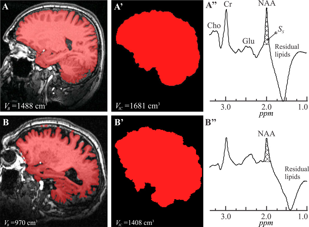
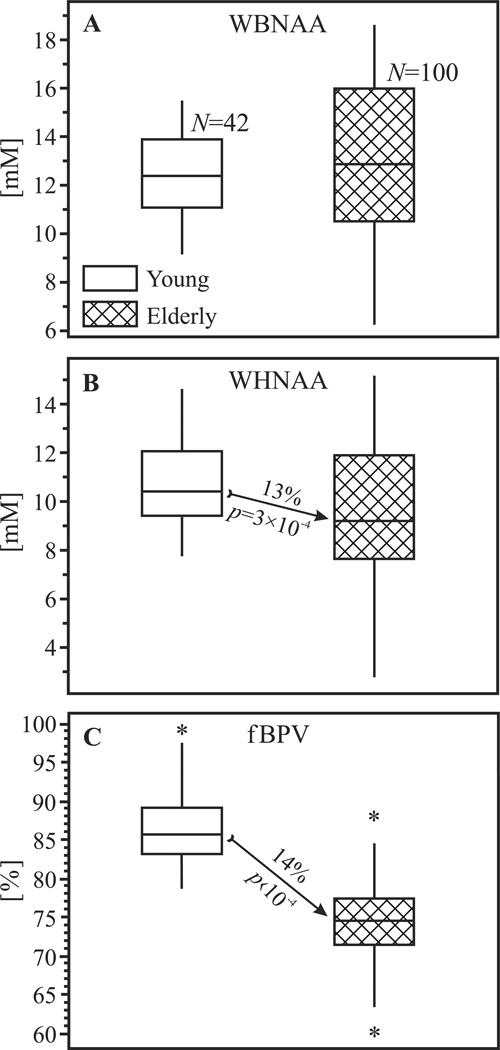
We hypothesize that normal aging implies neuronal durability, reflected by age-independent concentrations of their marker--the amino acid derivative N-acetylaspartate (NAA). To test this, we obtained the whole-brain and whole-head N-acetylaspartate concentrations (WBNAA and WHNAA) with proton magnetic resonance (MR) spectroscopy; and the fractional brain parenchyma volume (fBPV)--a metric of atrophy, by segmenting the magnetic resonance image (MRI) from 42 (18 male) healthy young (31.9 ± 5.8 years old) and 100 (64 male, 72.6 ± 7.3 years old) cognitively normal elderly. The 12.8 ± 1.9 mM WBNAA of the young was not significantly different from the 13.1 ± 3.1 mM in the elderly (p > 0.05). In contrast, both fBPV (87.3 ± 4.7% vs. 74.8 ± 4.8%) and WHNAA (11.1 ± 1.7 mM vs. 9.8 ± 2.4 mM) were significantly higher in the young (approximately 14%; p < 0.0001 for both). The similarity in mean WBNAA between 2 cohorts 4 decades of normal aging apart suggests that neuronal integrity is maintained across the lifespan. Clinically, WBNAA could be used as a marker for normal (hence, also abnormal) brain aging. In contrast, WHNAA and fBPV seem age-related suggesting that brain atrophy may occur without compromising the remaining tissue.
Copyright © 2012 Elsevier Inc. All rights reserved.
Conflict of interest statement
The authors state that there are no actual or potential conflicts of interest related to this paper.
Figures
References
-
- Army . Army Individual Test Battery Manual of Directions and Scoring. Washington DC: War Department, Adjutant General’s Office; 1944.
-
- Baslow MH. N-acetylaspartate in the vertebrate brain: metabolism and function. Neurochem Res. 2003;28:941–953. - PubMed
-
- Benarroch EE. N-acetylaspartate and N-acetylaspartylglutamate: neurobiology and clinical significance. Neurology. 2008;70:1353–1357. - PubMed
Publication types
MeSH terms
Substances
Grants and funding
LinkOut - more resources
Full Text Sources
Medical
