Review
. 2012 Mar;35(3):185-96.
doi: 10.1016/j.tins.2011.12.001. Epub 2012 Jan 12.

The vestibular system: multimodal integration and encoding of self-motion for motor control

Affiliations
Review

The vestibular system: multimodal integration and encoding of self-motion for motor control

Kathleen E Cullen. Trends Neurosci. 2012 Mar.

Abstract

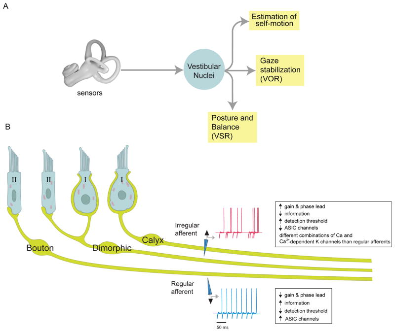
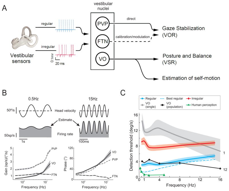
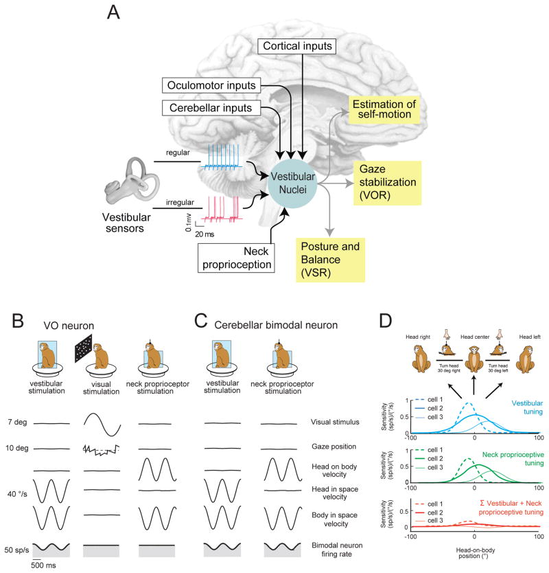
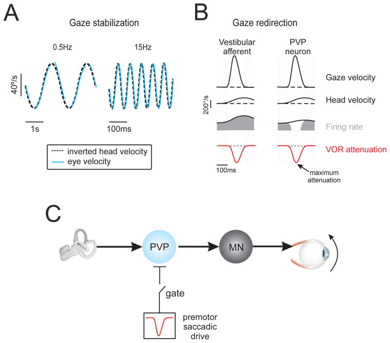
Understanding how sensory pathways transmit information under natural conditions remains a major goal in neuroscience. The vestibular system plays a vital role in everyday life, contributing to a wide range of functions from reflexes to the highest levels of voluntary behavior. Recent experiments establishing that vestibular (self-motion) processing is inherently multimodal also provide insight into a set of interrelated questions. What neural code is used to represent sensory information in vestibular pathways? How do the interactions between the organism and the environment shape encoding? How is self-motion information processing adjusted to meet the needs of specific tasks? This review highlights progress that has recently been made towards understanding how the brain encodes and processes self-motion to ensure accurate motor control.


Figures

Figure 1
Figure 1. Early Vestibular Processing and the Sensory Coding of Self-Motion: The Sensory Periphery
a) Vestibular signals from the labyrinth of the inner ear are transferred to the vestibular nuclei (VN) via the vestibular afferents of the VIII nerve. In turn, the VN projects to other brain areas to i) stabilize the visual axis of gaze via the vestibulo-ocular reflex, ii) control posture and balance, and iii) produce estimates of self-motion. b) Drawing of a regular afferent’s bouton ending contacting a type II hair cell (cell B), an irregular afferent’s calyx ending around a type I hair cell (cell C), and an irregular afferent contacting both types of hair cells [i.e. a dimorphic hair cell (cell D) also termed a D-irregular)]. Insets show example extracellular traces highlighting the difference in the resting discharge variability of regular (blue) and irregular (red) afferents. Abbreviations: acid-sensing (ASIC) conductances, ACIS.
Figure 2
Figure 2. Early Vestibular Processing and the Sensory Coding of Self-Motion: Central Neurons
a) Neurons in the vestibular nuclei that receive direct input from the vestibular afferents can be categorized into two main categories i) neurons that control and modulate the vestibulo-ocular reflex to ensure gaze stability during everyday life (i.e., PVPs and FTNs), and ii) neurons that control posture and balance, and also project to higher order structures involved in the estimation of self-motion (i.e., VO neurons). b) Firing rate response of an example VO neuron in the vestibular nucleus recorded in alert monkeys during sinusoidal head rotation at 0.5 and 15 Hz. Plots below show response gains averaged for populations of VO, PVP, and FTN neurons recorded over a wide range of frequencies of head rotation. Note, PVP neurons have relatively higher gains which increase more dramatically at higher frequencies. Side bands show +/-1 SEM. Data replotted from [32]. c) Average detection threshold values for regular (blue) and irregular (red) afferents, and VO neurons (gray) at different frequencies of sinusoidal head rotation in alert monkeys. Estimates of the information transmitted by a pooled population of 12 VO neurons (black) as well as human behavioral thresholds [37] are superimposed (green) for comparison. Side bands show +/- SEM. Data replotted from [13].
Figure 3
Figure 3. Multimodal integration within vestibular pathways
a) The vestibular nuclei (VN) receive direct input from multiple brain areas including: i) the vestibular afferents of the VIII nerve, ii) oculomotor areas of the brainstem, iii) the vestibular cerebellum, and iv) several areas of cortex [e.g., parietoinsular vestibular cortex (PIVC)], premotor areas 6, 6pa, somatosensory area 3a, and superior temporal cortex. b) VO neurons in the vestibular nuclei of the rhesus monkey are sensitive to vestibular stimulation, but are not well modulated by full field visual or neck proprioceptive stimulation [25, 44, 45, 55]. c,d) Neurons in the rostral fastigial nucleus of the vestibular cerebellum receive input from VO neurons. c. 50% of rostral fastigial neurons respond to neck proprioceptive (center) as well as vestibular (left) stimulation (i.e., bimodal neurons) [58]. d. When the head moves relative to body (as it would during a voluntary orienting head turn) the vestibular and dynamic neck proprioceptive inputs sum to produce complete response cancellation, consistent with these neurons’ encoding body motion. Vestibular (blue) and neck (green) turning curves are shown for 3 example neurons: cell 1 (dashed curve), cell 2 (solid thick curve), and cell 3 (solid thin curve). Note, for each cell responses to each modality sum linearly during combined stimulation such that bimodal neurons are not modulated during head-on-body motion (red curves). Thus, by combining their vestibular and neck related inputs, these neurons effectively encode body-in-space motion, rather than head-in-space motion. Data in (d) replotted with permission from [58].
Figure 4
Figure 4. The vestibulo-ocular reflex (VOR): compensatory response dynamics ensure stable gaze
a) The VOR is compensatory over a wide frequency range. Example eye and head velocity traces, during sinusoidal rotations of the head-on-body in the dark at 0.5 and 15 Hz. b) Single unit recording experiments in monkeys show that vestibular afferents encode the active head movements made during gaze shifts. However, neurons at the next stage of processing in the VOR pathways (i.e. PVP neurons) and resultant VOR are attenuated (red trace). The time course of the neuronal [34, 74] and VOR suppression [69] are comparable; response attenuation is maximal early in the gaze shift and progressively recovers to reach normal (i.e. compensatory) values near gaze-shift end. c) Mechanism underlying VOR suppression during gaze shifts. In addition to their input from the vestibular nerve, PVP neurons receive a strong inhibitory input from the premotor saccadic pathway, which effectively suppresses their activity during gaze shifts. In this way, VOR suppression is mediated by behaviorally-dependent gating of an inhibitory gaze command signal. Accordingly, during gaze shifts PVP neuron responses can be explained by the linear summation of their i) head velocity input and ii) this inhibitory saccadic drive.
Figure 5
Figure 5. Neural mechanism for the attenuation of vestibular reafference
To produce an active head movement, the brain sends a motor command to the neck muscle. The activation of the neck muscle moves the head, which in turn results in vestibular stimulation (i.e., vestibular reafference). In addition, the brain has access to an efference copy of the motor command and/or feedback from neck proprioceptors. a) Vestibular reafference is cancelled when neck proprioceptive feedback matches the expected sensory consequence of neck motor command (red shaded box: internal model followed by a putative matching operation). In this condition, a cancellation signal is sent to VO neurons in the vestibular nuclei. b) In the cerebellum-like structures of the mormyrid fish, the principals cells (Pc) receive an efference copy of the motor commands to the electric organ via parallel fibers, as well as afferent input from the electrosensory receptors. To remove predictable features of the sensory input (i.e., electrosensory reafference), anti-Hebbian plasticity at parallel fiber synapses generates “negative images” that act to cancel predictable patterns of electrosensory input [–87]. c) It remains to be determined whether a similar strategy is used by the mammalian cerebellum to selectively suppress vestibular reafference. Parallel fibers carry sensory as well as motor information to Purkinje cells (Pc), and climbing fibers are thought to encode a motor performance error signal (see review [26]). While climbing fiber activity paired in time with mossy fiber-parallel fiber activity is thought to weaken the associated parallel fiber synapse, a recent report suggest that instructive signals carried by parallel fiber activity alone may be sufficient to induce synaptic changes [94].

Similar articles

Cited by

References

    1. Angelaki DE, Cullen KE. Vestibular system: the many facets of a multimodal sense. Annual review of neuroscience. 2008;31:125–150. - PubMed
    1. Goldberg JM. Afferent diversity and the organization of central vestibular pathways. Experimental brain research. Experimentelle Hirnforschung. Experimentation cerebrale. 2000;130:277–297. - PMC - PubMed
    1. Eatock RA, Songer JE. Vestibular Hair Cells and Afferents: Two Channels for Head Motion Signals. Annual review of neuroscience. 2011;34:501–534. - PubMed
    1. Kalluri R, et al. Ion channels set spike timing regularity of Mammalian vestibular afferent neurons. Journal of neurophysiology. 2010;104:2034–2051. - PMC - PubMed
    1. Armand M, Minor LB. Relationship between time- and frequency-domain analyses of angular head movements in the squirrel monkey. Journal of computational neuroscience. 2001;11:217–239. - PubMed

Publication types