

Optical coherence tomography findings in autoimmune retinopathy
- PMID: 22245461
- PMCID: PMC3495560
- DOI: 10.1016/j.ajo.2011.09.012
Optical coherence tomography findings in autoimmune retinopathy
Abstract
Purpose: To report optical coherence tomography (OCT) features of patients with autoimmune retinopathy.
Design: Consecutive case series.
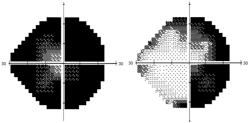
Method: Eight patients who presented with unexplained loss of central vision, visual field defects, and/or photopsia were diagnosed with autoimmune retinopathy based on clinical features, electroretinogram (ERG) findings, and serum antiretinal antibody analysis. All patients underwent OCT testing of the macula and nerve fiber layer (NFL).
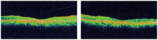
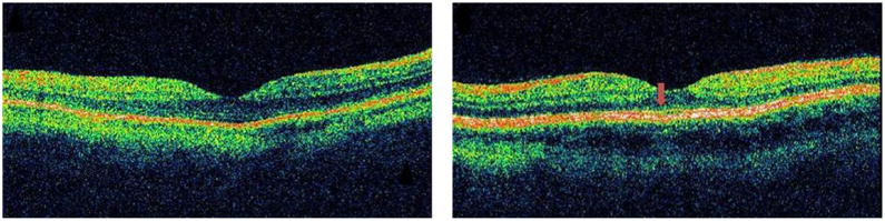
Results: Outer retinal abnormalities and/or decreased macular thickness on OCT were seen in all patients. Macular OCT showed reduced central macular and foveal thicknesses in 6 patients (mean thickness 143±30 μm and 131±29 μm respectively). In all but 1 patient, loss of the photoreceptor layer or disruption of the photoreceptor outer and inner segment junction was noted. Three patients showed only mild to moderate focal NFL loss.
Conclusions: Retinal atrophy and reduced macular thickness on OCT are predominant features in patients with autoimmune retinopathy. OCT provides objective measures of retinal damage and may offer clues toward understanding the mechanism of visual dysfunction and the diagnosis of autoimmune retinopathy.
Copyright © 2012 Elsevier Inc. All rights reserved.
Conflict of interest statement
All authors have completed and submitted the icmje form for disclosure of potential conflicts of Interest and none were reported.
Figures
References
-
- Ohguro H, Yokoi Y, Ohguro I, et al. Clinical and immunologic aspects of cancer-associated retinopathy. Am J Ophthalmol. 2004;137(6):1117–1119. - PubMed
-
- Weleber RG, Watzke RC, Shults WT, et al. Clinical and electrophysiologic characterization of paraneoplastic and autoimmune retinopathies associated with antienolase antibodies. Am J Ophthalmol. 2005;139(5):780–794. - PubMed
-
- Whitcup SM, Vistica BP, Milam AH, Nussenblatt RB, Gery I. Recoverin-associated retinopathy: a clinically and immunologically distinctive disease. Am J Ophthalmol. 1998;126(2):230–237. - PubMed
-
- Ohguro H, Ogawa K, Maeda T, Maeda A, Maruyama I. Cancer-associated retinopathy induced by both anti-recoverin and anti-hsc70 antibodies in vivo. Invest Ophthalmol Vis Sci. 1999;40(13):3160–3167. - PubMed
MeSH terms
Substances
Grants and funding
LinkOut - more resources
Full Text Sources
Medical
