. 2012 Apr;33(10):2961-70.
doi: 10.1016/j.biomaterials.2011.12.052. Epub 2012 Jan 14.

The resistance of breast cancer stem cells to conventional hyperthermia and their sensitivity to nanoparticle-mediated photothermal therapy

Affiliations

The resistance of breast cancer stem cells to conventional hyperthermia and their sensitivity to nanoparticle-mediated photothermal therapy

Andrew R Burke et al. Biomaterials. 2012 Apr.

Abstract

Breast tumors contain a small population of tumor initiating stem-like cells, termed breast cancer stem cells (BCSCs). These cells, which are refractory to chemotherapy and radiotherapy, are thought to persist following treatment and drive tumor recurrence. We examined whether BCSCs are similarly resistant to hyperthermic therapy, and whether nanoparticles could be used to overcome this resistance. Using a model of triple-negative breast cancer stem cells, we show that BCSCs are markedly resistant to traditional hyperthermia and become enriched in the surviving cell population following treatment. In contrast, BCSCs are sensitive to nanotube-mediated thermal treatment and lose their long-term proliferative capacity after nanotube-mediated thermal therapy. Moreover, use of this therapy in vivo promotes complete tumor regression and long-term survival of mice bearing cancer stem cell-driven breast tumors. Mechanistically, nanotube thermal therapy promotes rapid membrane permeabilization and necrosis of BCSCs. These data suggest that nanotube-mediated thermal treatment can simultaneously eliminate both the differentiated cells that constitute the bulk of a tumor and the BCSCs that drive tumor growth and recurrence.


Figures

Figure 1
Figure 1. Breast cancer stem cells are resistant to hyperthermic cell death and over-express HSP90
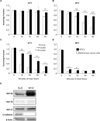
a–d, Relative viability of cells 24 hours after heat treatment. Cells were heated by water bath at 43°C (a), 45°C (b), 47°C (c) or 49°C (d) for 0–60 minutes. MTT absorbance values were normalized to the untreated condition (“0” minutes heat shock). Graphed are means (n=4) ± s.d. P-values correspond to the indicated pair-wise comparisons and were determined by post-hoc testing following an ANOVA that determined overall significance. (e) Expression levels of heat shock proteins (HSPs) 27, 70 and 90 were determined by Western blot. E-cadherin levels were used to confirm status of cell lines. β-actin served as a loading control. HSP90 is overexpressed ~4.5-fold in BCSCs relative to bulk breast cancer HMLER cells.
Figure 2
Figure 2. Sub-lethal hyperthermia enriches for the BCSC phenotype in bulk breast cancer cells
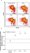
Changes in the CD44high/CD24low stem cell fraction of bulk breast cancer cells 24 hours after water bath heat shock at 43°, 45° or 47°C as determined by flow cytometry. These treatments killed 67.7, 71.3 and 82% of the starting cells by the time of analysis. a, Representative density dot plots show significant enrichment of CD44high/CD24low stem cells (indicated by black arrows) in the fraction of cells surviving heat treatment. b, Quantification of 3 experiments showing a significant enrichment of the CD44high/CD24low stem cell phenotype in viable cells 24 hours after treatment. Graphed are mean percent changes (n=3 ± s.d.) in the CD44high/CD24low cell fraction normalized to the Untreated condition (which is set as 1.0, i.e. “0 percent change”). Dashed lines indicate the 95% C.I. for the Untreated condition. All heat treatments led to significant increases in the stem cell fraction (p<0.0001) relative to Untreated. Significance was determined by ANOVA with post-hoc testing.
Figure 3
Figure 3. Stem and bulk breast cancer cells are equally sensitive to MWCNT-mediated thermal therapy
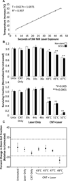
Breast cancer stem cells (BCSCs) and bulk breast cancer cells were rapidly heat treated to specific temperatures by the combination of MWCNTs and 3W NIR laser irradiation. NIR exposure ranged from 5–46 seconds. a, Change in temperature of a 50 µg/mL MWCNT suspension is linearly related (R2 = 0.997) to seconds of 3W NIR laser exposure. b, Relative viability of breast cancer cells 24 hours after MWCNT-mediated thermal therapy was determined by MTT. Graphed are means (n=4) ± s.d. normalized to the “Untreated” conditions. “CNT Only” describes samples that were mixed with MWCNTs but were not laser treated. “Laser Only” describes samples that were laser treated but did not contain MWCNTs. The indicated seconds of laser exposure in the “Laser Only” group correspond to the laser exposure times used in the “CNT + Laser” group. “CNT + Laser” describes samples that were heat shocked to the indicated final temperatures by the combination of 50 µg/mL MWCNTs and 3W laser radiation. P-values indicate significant differences for both cell types relative to the “Untreated” conditions. Statistical significance was determined by ANOVA and post-hoc testing. c, Bulk breast cancer cells were heat treated to the indicated final temperatures by nanotube-mediated thermal therapy. Changes in the CD44high/CD24low stem cell fraction were determined by flow cytometry. Quantification of 3 experiments showing no enrichment of the stem cell phenotype in viable cells 24 hours after treatment. “Laser Only” indicates samples that were laser treated but did not contain MWCNTs. “CNT Only” indicates samples that were mixed with 50 µg/mL MWCNTs but were not laser treated. Graphed are mean percent changes (n=3 ± s.d.) in the CD44high/CD24low cell fraction normalized to the “Untreated” condition (defined as “0 percent change”). Dashed lines indicate the 95% C.I. for the “Untreated” condition. Significance was determined by ANOVA with post-hoc testing.
Figure 4
Figure 4. Nanotube-mediated thermal therapy is more cytotoxic than water bath hyperthermia at equivalent exposure times and treatment temperatures
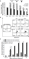
a, Relative viability of BCSCs cells following slow (“Slow ROTI”) or rapid (“Rapid ROTI”) induction of hyperthermia by water bath treatment or nanotube-mediated thermal treatments. Viability was assessed 24 hours after treatment. Graphed are mean (n=4) ± s.d. for each treatment group normalized to the appropriate Untreated condition. P-values correspond to the statistical differences between the NMTT treatment and ROTI treatments at a given temperature. Overall significance was determined by ANOVA with post-hoc tests for the pair-wise comparisons. (ROTI = rate of temperature increase; see Materials and Methods). b, Representative dot plots of BCSCs showing 7-AAD uptake and Annexin V labeling as a function of time post heat treatment. c, Quantification of total cell death (as determined by 7-AAD uptake) in both stem and bulk breast cancer cells following rapid ROTI water bath hyperthermia or NMTT to 53°C. Graph is representative of two independent experiments. Solid colors indicate cell type; pattern indicates form of heat treatment.
Figure 5
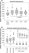
Figure 5. Nanotube-mediated thermal therapy but not water bath hyperthermia abrogates mammosphere-forming ability of BCSCs
BCSCs were heat shocked to 43–49°C by either rapid ROTI water bath hyperthermia (a) or NMTT (b) and then allowed to form mammospheres for 10 days. Graphed are median mammosphere diameters for each treatment group along with 25th and 75th percentiles. Outlier values are indicated by filled circles. At least 40 mammospheres were measured per treatment group. b, Inset provides detail on median mammosphere sizes formed after NMTT. Statistical differences were determined by ANOVA. **P<0.0001 relative to Untreated.
Figure 6
Figure 6. Long-term survival of mice bearing stem cell driven breast tumors following nanotube-mediated thermal therapy
Cohorts of mice (n=10 per group) bearing tumors derived from the breast cancer stem cell cell line were treated with NMTT (CNT + Laser) or control conditions (Untreated, CNT Only or Laser Only). Changes in tumor volume and overall survival were tracked for 45 days following treatment. Kaplan-Meier plot details the significant survival advantage of mice treated with NMTT relative to all other control groups (p<0.05).

References

    1. Kondo T, Setoguchi T, Taga T. Persistence of a small subpopulation of cancer stem-like cells in the C6 glioma cell line. Proc Natl Acad Sci U S A. 2004;101:781–786. - PMC - PubMed
    1. Eramo A, Lotti F, Sette G, Pilozzi E, Biffoni M, Di Virgilio A, et al. Identification and expansion of the tumorigenic lung cancer stem cell population. Cell Death Differ. 2008;15:504–514. - PubMed
    1. Prince ME, Sivanandan R, Kaczorowski A, Wolf GT, Kaplan MJ, Dalerba P, et al. Identification of a subpopulation of cells with cancer stem cell properties in head and neck squamous cell carcinoma. Proc Natl Acad Sci U S A. 2007;104:973–978. - PMC - PubMed
    1. Li C, Heidt DG, Dalerba P, Burant CF, Zhang L, Adsay V, et al. Identification of pancreatic cancer stem cells. Cancer Res. 2007;67:1030–1037. - PubMed
    1. Singh SK, Hawkins C, Clarke ID, Squire JA, Bayani J, Hide T, et al. Identification of human brain tumour initiating cells. Nature. 2004;432:396–401. - PubMed

Publication types

MeSH terms

Substances