Review
. 2012 Apr 15;302(8):H1546-62.
doi: 10.1152/ajpheart.00944.2011. Epub 2012 Jan 13.

New mechanisms of pulmonary arterial hypertension: role of Ca²⁺ signaling

Affiliations
Review

New mechanisms of pulmonary arterial hypertension: role of Ca²⁺ signaling

Frank K Kuhr et al. Am J Physiol Heart Circ Physiol. .

Abstract

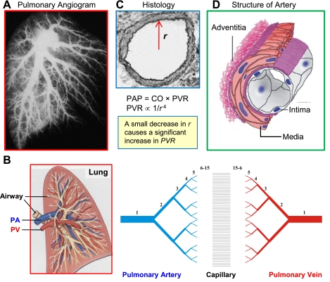
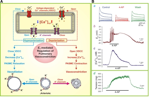
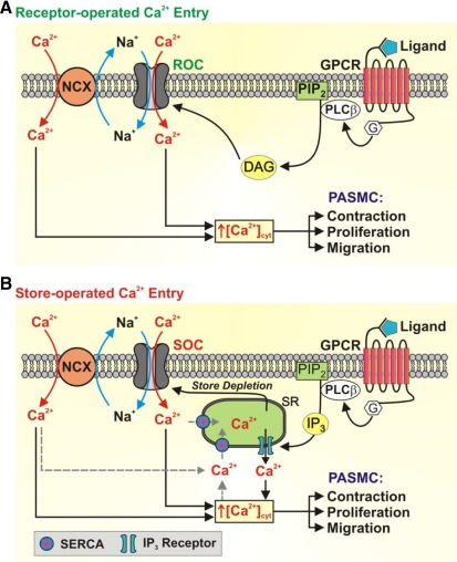
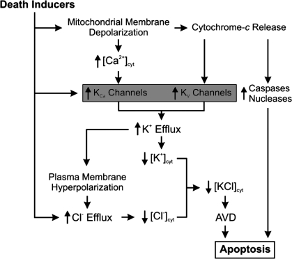
Pulmonary arterial hypertension (PAH) is a severe and progressive disease that usually culminates in right heart failure and death if left untreated. Although there have been substantial improvements in our understanding and significant advances in the management of this disease, there is a grim prognosis for patients in the advanced stages of PAH. A major cause of PAH is increased pulmonary vascular resistance, which results from sustained vasoconstriction, excessive pulmonary vascular remodeling, in situ thrombosis, and increased pulmonary vascular stiffness. In addition to other signal transduction pathways, Ca(2+) signaling in pulmonary artery smooth muscle cells (PASMCs) plays a central role in the development and progression of PAH because of its involvement in both vasoconstriction, through its pivotal effect of PASMC contraction, and vascular remodeling, through its stimulatory effect on PASMC proliferation. Altered expression, function, and regulation of ion channels and transporters in PASMCs contribute to an increased cytosolic Ca(2+) concentration and enhanced Ca(2+) signaling in patients with PAH. This review will focus on the potential pathogenic role of Ca(2+) mobilization, regulation, and signaling in the development and progression of PAH.


Figures

Fig. 1.
Fig. 1.
Structure and architecture of the pulmonary vasculature. A: pulmonary angiogram of the human lung showing the structure of the vasculature tree from large conducting vessels down to higher generation branches (arterioles). B: scheme of the pulmonary vasculature depicts the 15 orders of pulmonary arteries and veins with capillaries in between. C: cross section of pulmonary artery showing a small decrease in radius (r) or diameter can lead to a significant increase in pulmonary vascular resistance (PVR) and, as a result, elevation of pulmonary arterial pressure (PAP). D: schematic demonstrating the 3-layer structure of an artery from the lumen to the outside of the vessel. PA, pulmonary artery; PV, pulmonary vein; CO, cardiac output.
Fig. 2.
Fig. 2.
Morphological and functional changes of pulmonary arteries resulting from hypoxia. A: acute hypoxia induces pulmonary vasoconstriction (or decreased intraluminal diameter of pulmonary artery) leading to an increase in PVR with a corresponding increase in mean PAP (mPAP). The numbers shown in the box are the hypothetical prediction of the changes in mPAP before and after hypoxia based on the changes in pulmonary arterial diameter (top and middle). Arrows indicate the arterial branches that underwent significant pulmonary vasoconstriction during hypoxia. B: smaller arteries are also affected by hypoxia as indicated with vessel contraction in the smaller branches of the pulmonary tree. Images are reproduced with permission from Dawson (31).
Fig. 3.
Fig. 3.
Major causes for the elevated PVR in patients with pulmonary arterial hypertension (PAH): the effects of remodeling and corresponding changes in PVR. A: a significant loss of the small vessels of the pulmonary vasculature can be seen in an angiogram from a healthy (normal) patient versus a patient with PAH. Histological cross sections of small arteries reveal the extensive hypertrophy of the adventitia, media, and intima (or concentric pulmonary arterial remodeling) that occurs in patients with PAH [adapted by permission from BMJ Publishing Group Limited. Bedford et al. (8)]. B: list of the major causes leading to an increase PVR in patients with PAH. C: small arteries from a normal subject and 2 different patients with PAH show the change in architecture [adventitial and medial hypertrophy, intimal lesion, and intraluminal obliteration (top), as well as significant medial hypertrophy (bottom)] that occurs in the pulmonary vasculature causing elevated PVR. PASMC, pulmonary artery smooth muscle cell; PAEC, pulmonary artery endothelial cells.
Fig. 4.
Fig. 4.
Potential pathogenic mechanisms involved in PAH. Flow chart demonstrating the 4 major causes of elevated PVR and how they lead to PAH and eventually right heart failure.
Fig. 5.
Fig. 5.
An increase in cytosolic Ca2+ concentration ([Ca2+]cyt) in PASMCs is required for pulmonary vasoconstriction and plays an important role in cell proliferation and vascular remodeling. A: when the [Ca2+]cyt rises because of Ca2+ influx through different Ca2+ channels in the plasma membrane and Ca2+ mobilization from the intracellular stores [e.g., sarcoplasmic reticulum/endoplasmic reticulum (SR/ER)], Ca2+ binds calmodulin (CaM) which causes PASMC contraction by activating (via phosphorylation) myosin light chain (MLC) kinase (MLCK). Increased [Ca2+]cyt also activates CaM kinase (CaMK) and mitogen-activated protein kinase (MAPK), as well as other transcription factors [nuclear factor of activated T cells (NFAT), cAMP response element binding protein (CREB), activator protein-1 (AP-1), and NF-κB], to stimulate PASMC proliferation by propelling Ca2+-sensitive steps in the cell cycle progression. B: removal of extracellular Ca2+ (0 Ca) significantly inhibits vasoconstriction (determined by active tension) induced by 40 mM K+ (40 K) and phenylephrine (PE) in isolated rat pulmonary arterial rings (70). C: rat PASMC growth is significantly inhibited by chelation of extracellular Ca2+ with EGTA or by valinomycin (Val)-induced increase in K+ efflux (88). Cells are cultured in serum- and growth factor-contained media for 6 days in the absence of EGTA or Val (Cont), and the presence of 100 μM Val or 2 mM EGTA. MLCP, myosin light chain phosphatase; PAFB, pulmonary arterial fibroblasts; P, phosphorylated; G, M, S, phases of the cell cycle. **P < 0.01; ***P < 0.001.
Fig. 6.
Fig. 6.
The role of K+ channels in membrane depolarization and pulmonary vasoconstriction. A: when K+ channels are closed (or K+ channel expression is downregulated), the resulting membrane depolarization opens voltage-dependent Ca2+ channel (VDCC), promotes Ca2+ influx, increases [Ca2+]cyt, and causes vasoconstriction. When K+ channels are activated (or K+ channel gene expression is upregulated), the resulting membrane hyperpolarization closes VDCC, inhibits agonist-mediated Ca2+ influx and causes vasodilation. Adapted from Jackson (57) and Makino et al. (67). B: representative recordings showing whole cell K+ currents (a), membrane potential (Em, b), and [Ca2+]cyt in rat PASMCs before, during, and after extracellular application of the KV channel blocker 4-aminopyridine (4-AP; 5 mM). A representative recording of tension measurement in an isolated mouse pulmonary arterial ring before, during and after treatment with 4-AP (d). Wash, washout. Reproduced from Yuan (137) with permission.
Fig. 7.
Fig. 7.
Receptor-mediated increase in [Ca2+]cyt in PASMC via receptor-operated Ca2+ entry (ROCE) and store-operated Ca2+ entry (SOCE). A: upon binding of ligands to membrane receptors [such as G protein-coupled receptor (GPRC) and receptor tyrosine kinase (RTK)], PLC is activated leading to the production of diacylglycerol (DAG) and inositol-1,4,5-trisphosphate (IP3). Receptor-operated Ca2+ (ROC) channels are activated by DAG. B: store-operated Ca2+ (SOC) channels are activated as a result of store depletion resulting from IP3-mediated Ca2+ release from the SR. The opening of ROC and SOC results in an influx of not only Ca2+ but also Na+ [canonical transient receptor potential (TRPC) channels are permeable to Ca2+ and Na+]. The locally increased [Na+] activates the reverse mode of the Na+/Ca2+ exchanger (NCX), which contributes to the increased [Ca2+]cyt and PASMC contraction, proliferation, and migration. PIP2, phosphatidylinositol 4,5-bisphosphate; SERCA, sarco(endo)plasmic reticulum Ca2+ ATPase; G, G protein. Adapted from Song et al. (109).
Fig. 8.
Fig. 8.
The role of stromal-interacting molecule (STIM) and Orai in SOCE. The EF-hand domain in the NH2-terminus of STIM acts as a Ca2+ sensor and binds Ca2+ (red circles) when the SR/ER Ca2+ store is full or the intracellularly stored [Ca2+] in the SR/ER is around 1 mM. IP3-mediated Ca2+ store depletion causes Ca2+ to unbind from the low-affinity EF-hand domain of STIM and results in the oligomerization of Ca2+-depleted STIM dimers and their translocation to SR/ER-plasma membrane junctions. STIM accumulation in the vicinity of Orai channels induces Orai channels to open, allowing an influx of Ca2+. TKR, tyrosine kinase receptor; IP3R, IP3 receptor; N, NH2 terminal; PM, plasma membrane. Modified from Cahalan (20) with permission from Macmillan Publishers Ltd.
Fig. 9.
Fig. 9.
The pathogenic role of TRPC6 and NCX in idiopathic PAH (IPAH). A: genetic mutations and/or environmental stimulation leads to upregulation of TRPC6 resulting in an increased number of ROC and SOC channels in the plasma membrane and enhanced ROCE and SOCE. The enhanced ROCE/SOCE leads to not only increased [Ca2+]cyt but also to locally increased cytosolic Na+ concentration and activation of the reverse mode of NCX. NCX is also upregulated in patients with IPAH. These mechanisms contribute to increased [Ca2+]cyt and pulmonary vascular remodeling. B and C: representative currents (I) elicited by a ramp depolarization from −100 to +100 mV and [Ca2+]cyt in normal or control PASMCs and IPAH-PASMCs before and after treatment with 1-oleoyl-2-acetyl-sn-glycerol (OAG), a membrane permeable DAG analog. B and C reproduced from Yu et al. (133) with permission.
Fig. 10.
Fig. 10.
Functional coupling of TRPC channels and NCX in caveolae in PASMCs. A: electron micrograph (EM) images showing the increased number of caveolae (indicated by arrowheads) in PASMCs from patients with IPAH vs. PASMCs from normal subjects (Nor). Western blots demonstrate the differential expression levels of caveolin (Cav)-1, Cav-2, and Cav-3 in normal and IPAH PASMCs. Reproduced from Patel et al. (85) with permission. B: schematic diagram depicting the Cav-1 binding domains of TRPC channels and how the colocalization of NCX, TRPC, and membrane receptors, such as GPCR, in caveolae allows functional interactions between the components and enhances Ca2+ signaling. CBM, Cav-1-binding motif; PBD, protein 4-binding motif; CDS, caveolin-scaffolding domain.
Fig. 11.
Fig. 11.
Decreased expression of K+ channels contribute to the development of PAH by increasing [Ca2+]cyt and inhibiting apoptosis in PASMCs. Flow chart depicting how downregulation of K+ channels causes membrane depolarization and increased [Ca2+]cyt through VDCC, leading to pulmonary vasoconstriction and PAH and how decreased K+ efflux leads to reducing apoptotic volume decrease (AVD) and apoptosis in PASMCs from patients with PAH. [K+]cyt, cytosolic K+ concentration.
Fig. 12.
Fig. 12.
The role of activity of K+ channels in AVD and apoptosis in PASMCs. In addition to regulating membrane potential, the activity of K+ channels also contributes to the regulation of AVD, an early hallmark of apoptosis. Decreased expression of K+ channels in PASMCs from patients with IPAH leads to decreased apoptosis. KCa, Ca2+-activaed K+ channel; KV, voltage-gated K+ channel; [KCl]cyt, cytosolic KCl concentration; [Cl], cytosolic Cl concentration.
Fig. 13.
Fig. 13.
The positive-feedback loop hypothesis of Ca2+ signaling in PAH. Multiple Ca+ channels (ROC, SOC, VDCC, TRPC) contribute to regulating the [Ca2+]cyt in PASMC (A). An increase in [Ca2+]cyt, because of increased expression and/or function of Ca+ channels (or transporters), can lead to the activation of transcription factors and signaling pathways in a positive-feedback loop, which leads to a greater and persistent influx of Ca2+. The activated signaling pathways result in cross talk among many different genetic and signaling pathways and an interaction network of multiple genes and proteins (B). This ultimately leads to the processes (contraction, migration, imbalanced ratio of proliferation to apoptosis, misguided differentiation, dedifferentiation, transdifferentiation, partial reprogramming) in all cell types involved in the pulmonary vascular wall, which can contribute to the concentric pulmonary vascular remodeling (C) and, ultimately, PAH.

References

    1. Angermann JE, Sanguinetti AR, Kenyon JL, Leblanc N, Greenwood IA. Mechanism of the inhibition of Ca2+-activated Cl currents by phosphorylation in pulmonary arterial smooth muscle cells. J Gen Physiol 128: 73–87, 2006 - PMC - PubMed
    1. Archer S, Rich S. Primary pulmonary hypertension: a vascular biology and translational research “Work in progress”. Circulation 102: 2781–2791, 2000 - PubMed
    1. Archer SL, Souil E, Dinh-Xuan AT, Schremmer B, Mercier JC, El Yaagoubi A, Nguyen-Huu L, Reeve HL, Hampl V. Molecular identification of the role of voltage-gated K+ channels, Kv1.5 and Kv21, in hypoxic pulmonary vasoconstriction and control of resting membrane potential in rat pulmonary artery myocytes. J Clin Invest 101: 2319–2330, 1998 - PMC - PubMed
    1. Archer SL, Wu XC, Thebaud B, Nsair A, Bonnet S, Tyrrell B, McMurtry MS, Hashimoto K, Harry G, Michelakis ED. Preferential expression and function of voltage-gated, O2-sensitive K+ channels in resistance pulmonary arteries explains regional heterogeneity in hypoxic pulmonary vasoconstriction: ionic diversity in smooth muscle cells. Circ Res 95: 308–318, 2004 - PubMed
    1. Atkinson NS, Robertson GA, Ganetzky B. A component of calcium-activated potassium channels encoded by the Drosophila slo locus. Science 253: 551–555, 1991 - PubMed

Publication types

MeSH terms