

Multicenter assessment of the reproducibility of volumetric radiofrequency-based intravascular ultrasound measurements in coronary lesions that were consecutively stented
- PMID: 22246064
- PMCID: PMC3485535
- DOI: 10.1007/s10554-012-0011-y
Multicenter assessment of the reproducibility of volumetric radiofrequency-based intravascular ultrasound measurements in coronary lesions that were consecutively stented
Abstract
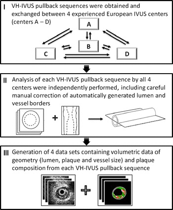
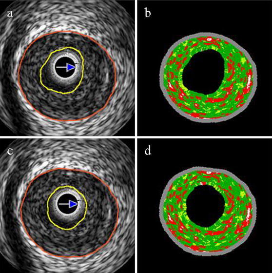
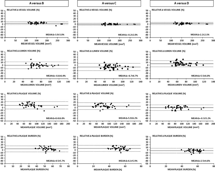
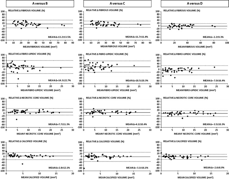
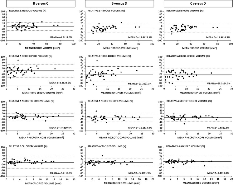
To assess in a multicenter design the between-center reproducibility of volumetric virtual histology intravascular ultrasound (VH-IVUS) measurements with a semi-automated, computer-assisted contour detection system in coronary lesions that were consecutively stented. To evaluate the reproducibility of volumetric VH-IVUS measurements, experienced analysts of 4 European IVUS centers performed independent analyses (in total 8,052 cross-sectional analyses) to obtain volumetric data of 40 coronary segments (length 20.0 ± 0.3 mm) from target lesions prior to percutaneous intervention that were performed in the setting of stable (65%) or unstable angina pectoris (35%). Geometric and compositional VH-IVUS measurements were highly correlated for the different comparisons. Overall intraclass correlation for vessel, lumen, plaque volume and plaque burden was 0.99, 0.92, 0.96, and 0.83, respectively; for fibrous, fibro-lipidic, necrotic core and calcified volumes overall intraclass correlation was 0.96, 0.94, 0.98, and 0.99, respectively. Nevertheless, significant differences for both geometrical and compositional measurements were seen. Of the plaque components, fibrous tissue and necrotic core showed on average the highest measurement reproducibility. A central analysis for VH-IVUS multicenter studies of lesions prior to PCI should be pursued. Moreover, it may be problematical to pool VH-IVUS data of individual trials analyzed by independent centers.
Figures

Similar articles
-
Between-centre reproducibility of volumetric intravascular ultrasound radiofrequency-based analyses in mild-to-moderate coronary atherosclerosis: an international multicentre study.EuroIntervention. 2010 Apr;5(8):925-31. EuroIntervention. 2010. PMID: 20542777
-
Automatic quantification and characterization of coronary atherosclerosis with computed tomography coronary angiography: cross-correlation with intravascular ultrasound virtual histology.Int J Cardiovasc Imaging. 2013 Jun;29(5):1177-90. doi: 10.1007/s10554-013-0194-x. Epub 2013 Feb 16. Int J Cardiovasc Imaging. 2013. PMID: 23417447
-
Reproducibility of Shin's method for necrotic core and calcium content in atherosclerotic coronary lesions treated with bioresorbable everolimus-eluting vascular scaffolds using volumetric intravascular ultrasound radiofrequency-based analysis.Int J Cardiovasc Imaging. 2012 Jan;28(1):43-9. doi: 10.1007/s10554-010-9779-9. Epub 2011 Jan 13. Int J Cardiovasc Imaging. 2012. PMID: 21229393
-
Multimodality imaging of attenuated plaque using grayscale and virtual histology intravascular ultrasound and optical coherent tomography.Catheter Cardiovasc Interv. 2016 Jul;88(1):E1-E11. doi: 10.1002/ccd.25786. Epub 2016 May 9. Catheter Cardiovasc Interv. 2016. PMID: 25511369
-
Clinical expert consensus document on intravascular ultrasound from the Japanese Association of Cardiovascular Intervention and Therapeutics (2021).Cardiovasc Interv Ther. 2022 Jan;37(1):40-51. doi: 10.1007/s12928-021-00824-0. Epub 2021 Nov 12. Cardiovasc Interv Ther. 2022. PMID: 34767160 Free PMC article. Review.
Cited by
-
Cardiovascular imaging 2012 in the International Journal of Cardiovascular Imaging.Int J Cardiovasc Imaging. 2013 Apr;29(4):725-36. doi: 10.1007/s10554-013-0216-8. Int J Cardiovasc Imaging. 2013. PMID: 23589003 Review. No abstract available.
References
-
- Garcia-Garcia HM, Mintz GS, Lerman A, Vince DG, Margolis MP, van Es GA, Morel MA, Nair A, Virmani R, Burke AP, Stone GW, Serruys PW. Tissue characterisation using intravascular radiofrequency data analysis: recommendations for acquisition, analysis, interpretation and reporting. Eurointervention. 2009;5(2):177–189. doi: 10.4244/EIJV5I2A29. - DOI - PubMed
-
- Hartmann M, Mattern ES, Huisman J, van Houwelingen GK, de Man FH, Stoel MG, Danse PW, Louwerenburg HW, von Birgelen C. Reproducibility of volumetric intravascular ultrasound radiofrequency-based analysis of coronary plaque composition in vivo. Int J Cardiovasc Imaging. 2009;25(1):13–23. doi: 10.1007/s10554-008-9338-9. - DOI - PMC - PubMed
-
- Mintz GS, Nissen SE, Anderson WD, Bailey SR, Erbel R, Fitzgerald PJ, Pinto FJ, Rosenfield K, Siegel RJ, Tuzcu EM, Yock PG. American college of cardiology clinical expert consensus document on standards for acquisition, measurement and reporting of intravascular ultrasound studies (IVUS). A report of the american college of cardiology task force on clinical expert consensus documents. J Am Coll Cardiol. 2001;37(5):1478–1492. doi: 10.1016/S0735-1097(01)01175-5. - DOI - PubMed
Publication types
MeSH terms
LinkOut - more resources
Full Text Sources
Medical
Miscellaneous
