. 2012 Mar 7;31(5):1266-78.
doi: 10.1038/emboj.2011.498. Epub 2012 Jan 13.

A DR4:tBID axis drives the p53 apoptotic response by promoting oligomerization of poised BAX

Affiliations

A DR4:tBID axis drives the p53 apoptotic response by promoting oligomerization of poised BAX

Ryan E Henry et al. EMBO J. .

Abstract

The cellular response to p53 activation varies greatly in a stimulus- and cell type-specific manner. Dissecting the molecular mechanisms defining these cell fate choices will assist the development of effective p53-based cancer therapies and also illuminate fundamental processes by which gene networks control cellular behaviour. Using an experimental system wherein stimulus-specific p53 responses are elicited by non-genotoxic versus genotoxic agents, we discovered a novel mechanism that determines whether cells undergo proliferation arrest or cell death. Strikingly, we observe that key mediators of cell-cycle arrest (p21, 14-3-3σ) and apoptosis (PUMA, BAX) are equally activated regardless of outcome. In fact, arresting cells display strong translocation of PUMA and BAX to the mitochondria, yet fail to release cytochrome C or activate caspases. Surprisingly, the key differential events in apoptotic cells are p53-dependent activation of the DR4 death receptor pathway, caspase 8-mediated cleavage of BID, and BID-dependent activation of poised BAX at the mitochondria. These results reveal a previously unappreciated role for DR4 and the extrinsic apoptotic pathway in cell fate choice following p53 activation.


Conflict of interest statement

The authors declare that they have no conflict of interest.

Figures

Figure 1
Figure 1
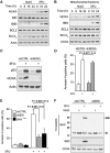
Stimulus-specific responses to p53 activation are defined by differential BAX oligomerization. (A) HCT116 cells undergo p53-dependent apoptosis in response to 5FU, but not Nut3. Cells were treated with Nut3 or 5FU for the indicated times, stained with Annexin-V-FITC and analysed by flow cytometry. Isogenic p53-null HCT116 cells are denoted as p53−/−. Data shown are the mean±s.d. of at least three independent experiments. (B) Immunoblot analysis reveals that several genes mediating cell-cycle arrest and apoptosis are expressed equally in response to Nut3 or 5FU treatment. Casp3* denotes the p19/17 doublet indicative of caspase 3 activation. (C) Nut3-treated cells accumulate as much mitochondrial PUMA and BAX as do 5FU-treated cells. COX4 is a mitochondrial protein serving as a loading control. HCT116 cells were treated as in (B) before preparation of mitochondrial fractions (see Supplementary Figure S1 for fractionation controls). (D) BAX oligomerization is observed only in 5FU-treated cells. Filled circles on the right represent the predicted migration of BAX mono-, di-, tri-, and tetramers. (E) Stimulus-specific activation of the BAX protein was detected by immunoprecipitation with the conformer-specific 6A7 antibody followed by western blot. Figure source data can be found in Supplementary data.
Figure 2
Figure 2
BID cleavage is stimulus-specific and required for BAX oligomerization and apoptosis. (A) NOXA and tBID proteins accumulate in 5FU-treated, but not in Nut3-treated cells, whereas BCL2 and BCL-XL levels are similar between treatments. (B) Levels of mitochondria-localized NOXA and tBID increase following 5FU, but not Nut3, whereas the levels of BCL2 and BCL-XL remain similar between treatments. The COX4 panel is the same as in Figure 1C. (C) Immunoblot assessment of NOXA knockdown. (D) Apoptotic index assays show no significant effect of NOXA knockdown on 5FU-induced apoptosis. Data shown are the mean±s.d. (E) Apoptotic index assays indicate that BID knockdown abrogates 5FU-induced apoptosis as effectively as BAX knockout. Data shown are the mean±s.d. (F) Western blot analysis of BAX dimerization in Nut3 and 5FU-treated cells expressing shCTRL or shBID. BID depletion prevents BAX dimerization in response to 5FU treatment. P-values shown in (D, E) were calculated by Student's t-test (unpaired, two-tailed). Figure source data can be found in Supplementary data.
Figure 3
Figure 3
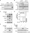
Caspase 8 activation is stimulus-specific, p53-dependent and required for BID cleavage and BAX activation. (A) Caspase 8 activation is stimulus-specific (FL) indicates full-length caspase 8 (55–57 kDa), Casp8* indicates the 43–41 kDa cleavage products. (B) 5FU-induced caspase 8 activation and BID cleavage in HCT116 cells are p53-dependent events. (C) Caspase 8 knockdown prevents caspase 3 activation and PARP cleavage upon 5FU treatment. White line indicates cropping of intervening lanes from the same blot. (D) Apoptotic index assays show that caspase 8 is required for 5FU-induced apoptosis. Data shown are the mean±s.d. P-values were calculated by Student's t-test (unpaired, two-tailed). (E) BID cleavage in 5FU-treated cells requires caspase 8. (F) Immunoblot analysis of 6A7 immunoprecipitates indicates that BAX activation following 5FU treatment is both caspase 8- and BID-dependent. Figure source data can be found in Supplementary data.
Figure 4
Figure 4
Stimulus-specific activation of the extrinsic apoptotic pathway is mediated by FADD. (A) Caspase 3 activation requires the adaptor protein FADD, not TRADD. (B) Apoptotic index assays demonstrate that FADD, not TRADD, is required for full induction of apoptosis upon 5FU treatment. Data shown are the mean±s.d. P-values were calculated by Student's t-test (unpaired, two-tailed). (C) Immunoblot analysis of 6A7 immunoprecipitates indicates that BAX activation following 5FU treatment is FADD-dependent. Figure source data can be found in Supplementary data.
Figure 5
Figure 5
DR4 is required for stimulus-specific BAX activation and apoptosis. (A) Immunoblots show that total protein levels of the DR4 and FAS death receptors, but not DR5, are higher in 5FU-treated cells than in those treated with Nut3. (*) Indicates a non-specific band. (B) Nut3 fails to induce DR4 cell surface expression as measured by flow cytometry. Nut3 and 5FU induce DR5 and FAS cell surface localization equally. Data shown are representative of a least three independent experiments. (C) Apoptotic index assays show that DR4, but not FAS, is required for 5FU-induced apoptosis (see Supplementary Figure S6 for western blot analysis of FAS knockdown). Data shown are the mean±s.d. P-values were calculated by Student's t-test (unpaired, two-tailed). (D) Immunoblot analysis of 6A7 immunoprecipitates indicates that BAX activation following 5FU treatment is DR4-dependent. Figure source data can be found in Supplementary data.
Figure 6
Figure 6
Stimulus-specific stabilization of the DR4 mRNA. (A) Q-RT–PCR analysis reveals that the DR4 mRNA, but not the p21 mRNA, accumulates more strongly in 5FU-treated cells. (B) ChIP analysis of the DR4 and p21 loci show increased levels of initiating (S5P-RNAPII) and elongating (S2P-RNAPII) forms of RNAPII in response to p53 activation by both Nut3 and 5FU. Grey regions represent the transcribed region of each locus, arrows indicate transcription start sites, black boxes represent exons, and black dashes indicate the position of PCR amplicons used for analysis of ChIP-enriched DNA. (C) Schematic of experimental procedure for DR4 mRNA half-life determination (top). Following 12 h of DMSO, Nut3 or 5FU treatment of HCT116 cells of different p53 status, RNA synthesis was halted by ActD and the levels of DR4, NOXA and MDM2 mRNA levels were measured over the time, revealing that 5FU treatment leads to stabilization of the DR4 and NOXA mRNAs in a p53-independent fashion. The numbers to the right indicate the mRNA half-lives for the corresponding mRNAs in HCT116 p53+/+ cells treated with DMSO (black) or 5FU (blue). A table with all half-life measurements can be found in Supplementary Figure S7.
Figure 7
Figure 7
The DR4/FADD/caspase 8/tBID axis also defines cell fate choice in the H460 lung cancer cell line. (A) Western blot analysis of H460 cells shows that both Nut3 and 5FU activate p53 effectively, yet only 5FU leads to caspase 8 activation, tBID formation and caspase 3 activation. Differential cell fate choice correlates with higher levels of DR4 at late time points after 5FU treatment. (B) Q-RT–PCR analysis shows that the DR4 mRNA accumulates only in 5FU-treated H460 cells. (C) Knockdown of BID, caspase 8, DR4, or FADD impairs both tBID formation and activation of the executioner caspase 3. Immunoblot assessment of knockdown efficiency in H460 cells can be found in Supplementary Figure S8. (D) Apoptotic index assays reveal that BID, caspase 8, DR4, and FADD are all required for efficient induction of apoptosis upon 5FU treatment. Data shown are the mean±s.d. The indicated P-values represent the statistical significance of the 5FU-treated value for the respective knockdown line relative to the CTRL shRNA value and were calculated by Student's t-test (unpaired, two-tailed). Figure source data can be found in Supplementary data.
Figure 8
Figure 8
Model of stimulus-specific configurations of the p53 network defining cell fate choice in response to Nutlin-3 and 5FU. See Discussion for details.

Similar articles

Cited by

References

    1. Beckerman R, Donner AJ, Mattia M, Peart MJ, Manley JL, Espinosa JM, Prives C (2009) A role for Chk1 in blocking transcriptional elongation of p21 RNA during the S-phase checkpoint. Genes Dev 23: 1364–1377 - PMC - PubMed
    1. Brown CJ, Lain S, Verma CS, Fersht AR, Lane DP (2009) Awakening guardian angels: drugging the p53 pathway. Nat Rev Cancer 9: 862–873 - PubMed
    1. Bunz F, Dutriaux A, Lengauer C, Waldman T, Zhou S, Brown JP, Sedivy JM, Kinzler KW, Vogelstein B (1998) Requirement for p53 and p21 to sustain G2 arrest after DNA damage. Science 282: 1497–1501 - PubMed
    1. Bunz F, Hwang PM, Torrance C, Waldman T, Zhang Y, Dillehay L, Williams J, Lengauer C, Kinzler KW, Vogelstein B (1999) Disruption of p53 in human cancer cells alters the responses to therapeutic agents. J Clin Invest 104: 263–269 - PMC - PubMed
    1. Burns TF, Bernhard EJ, El-Deiry WS (2001) Tissue specific expression of p53 target genes suggests a key role for KILLER/DR5 in p53-dependent apoptosis in vivo. Oncogene 20: 4601–4612 - PubMed

Publication types

MeSH terms

Substances