

Computerized analysis of mammographic parenchymal patterns on a large clinical dataset of full-field digital mammograms: robustness study with two high-risk datasets
- PMID: 22246204
- PMCID: PMC3447101
- DOI: 10.1007/s10278-012-9452-z
Computerized analysis of mammographic parenchymal patterns on a large clinical dataset of full-field digital mammograms: robustness study with two high-risk datasets
Abstract
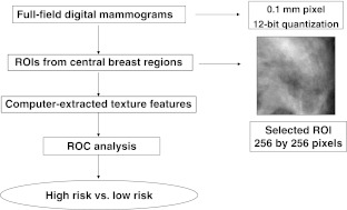
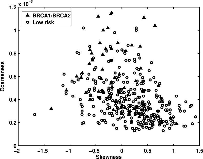
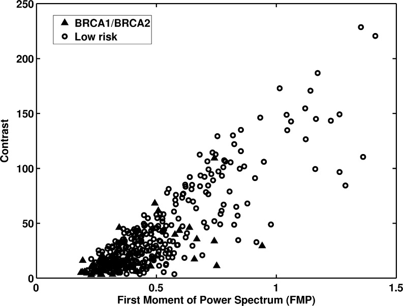
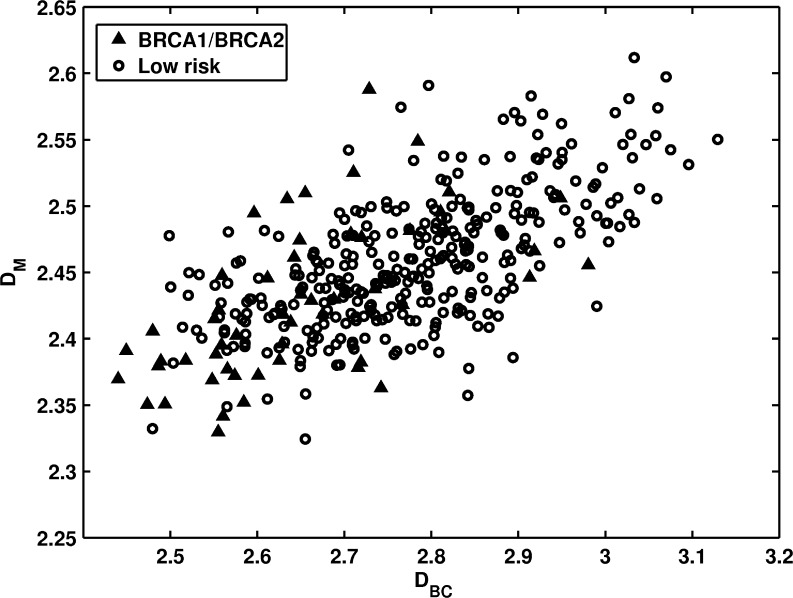
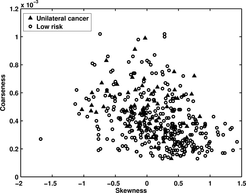
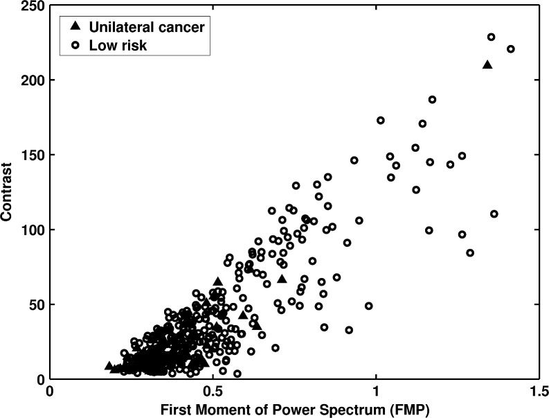
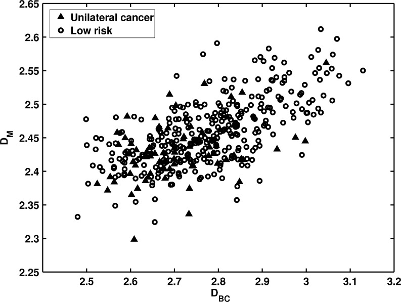
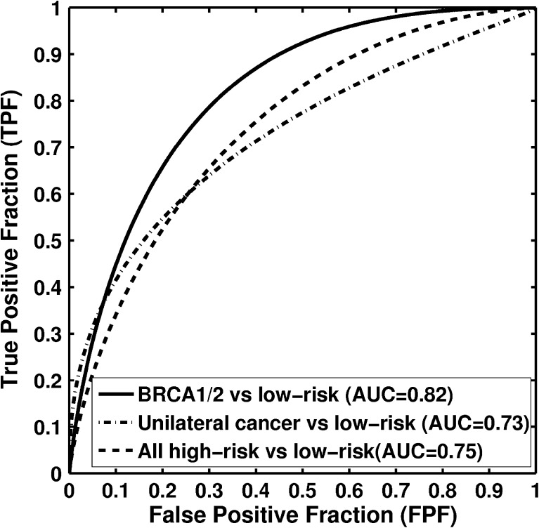
The purpose of this study was to demonstrate the robustness of our prior computerized texture analysis method for breast cancer risk assessment, which was developed initially on a limited dataset of screen-film mammograms. This current study investigated the robustness by (1) evaluating on a large clinical dataset, (2) using full-field digital mammograms (FFDM) as opposed to screen-film mammography, and (3) incorporating analyses over two types of high-risk patient sets, as well as patients at low risk for breast cancer. The evaluation included the analyses on the parenchymal patterns of women at high risk of developing of breast cancer, including both BRCA1/2 gene mutation carriers and unilateral cancer patients, and of women at low risk of developing breast cancer. A total of 456 cases, including 53 women with BRCA1/2 gene mutations, 75 women with unilateral cancer, and 328 low-risk women, were retrospectively collected under an institutional review board approved protocol. Regions-of-interest (ROIs), were manually selected from the central breast region immediately behind the nipple. These ROIs were subsequently used in computerized feature extraction to characterize the mammographic parenchymal patterns in the images. Receiver operating characteristic analysis was used to assess the performance of the computerized texture features in the task of distinguishing between high-risk and low-risk subjects. In a round robin evaluation on the FFDM dataset with Bayesian artificial neural network analysis, AUC values of 0.82 (95% confidence interval [0.75, 0.88]) and 0.73 (95% confidence interval [0.67, 0.78]) were obtained between BRCA1/2 gene mutation carriers and low-risk women, and between unilateral cancer and low-risk women, respectively. These results from computerized texture analysis on digital mammograms demonstrated that high-risk and low-risk women have different mammographic parenchymal patterns. On this large clinical dataset, we validated our methods for quantitative analyses of mammographic patterns on FFDM, statistically demonstrating again that women at high risk tend to have dense breasts with coarse and low-contrast texture patterns.
Figures
References
-
- Breast Imaging Reporting and Data System (BI-RADS) 4. Reston, Va: American College of Radiology; 2003.
-
- Wolfe JN. Breast patterns as an index of risk for developing breast cancer. Am J Roentgenol. 1976;126:1130–1139. - PubMed
-
- Brisson J, Diorio C, Mâsse B. Wolfe’s parenchymal pattern and percentage of the breast with mammographic densities: redundant or complementary classifications? Cancer Epidemiol Biomarkers Prev. 2003;12:728–732. - PubMed
Publication types
MeSH terms
Grants and funding
LinkOut - more resources
Full Text Sources
Medical
Miscellaneous
