

Increased PARP-1 association with DNA in alkylation damaged, PARP-inhibited mouse fibroblasts
- PMID: 22246237
- PMCID: PMC3307909
- DOI: 10.1158/1541-7786.MCR-11-0477
Increased PARP-1 association with DNA in alkylation damaged, PARP-inhibited mouse fibroblasts
Abstract
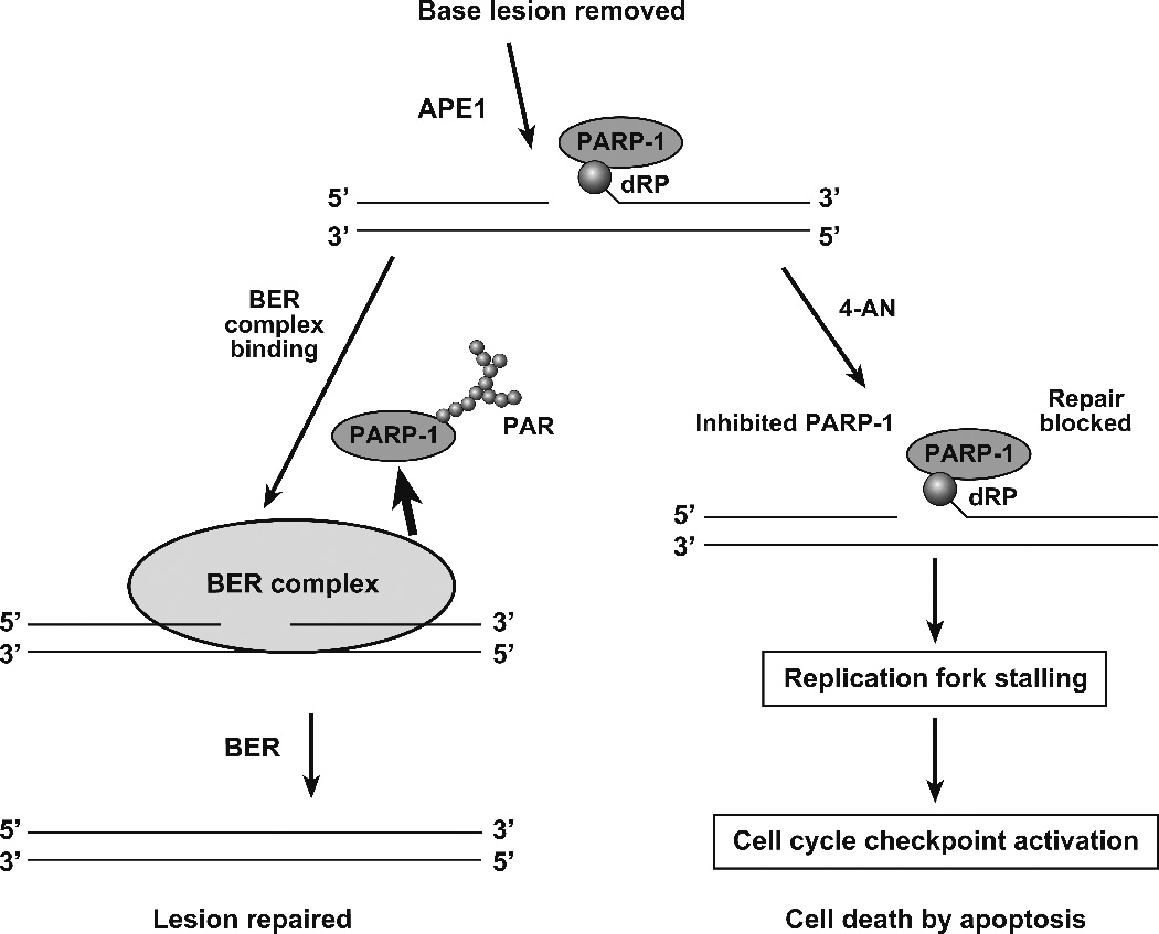
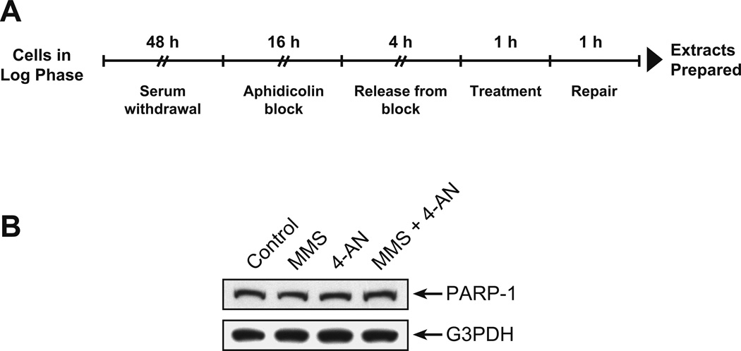
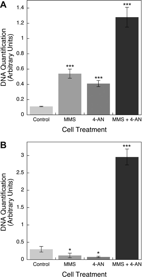
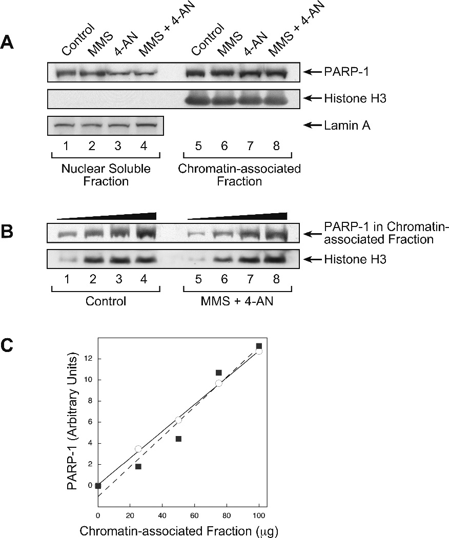
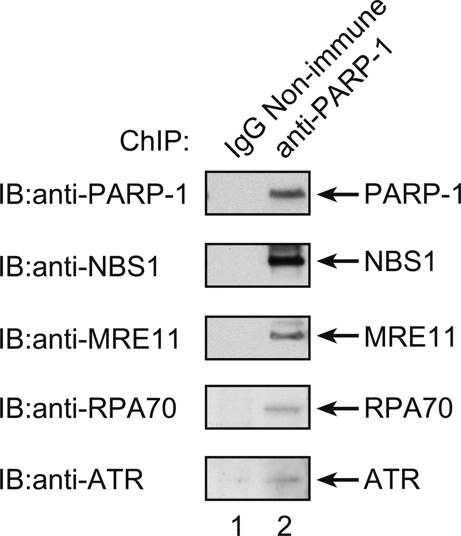
Treatment of base excision repair-proficient mouse fibroblasts with the DNA alkylating agent methyl methanesulfonate (MMS) and a small molecule inhibitor of PARP-1 results in a striking cell killing phenotype, as previously reported. Earlier studies showed that the mechanism of cell death is apoptosis and requires DNA replication, expression of PARP-1, and an intact S-phase checkpoint cell signaling system. It is proposed that activity-inhibited PARP-1 becomes immobilized at DNA repair intermediates, and that this blocks DNA repair and interferes with DNA replication, eventually promoting an S-phase checkpoint and G(2)-M block. Here we report studies designed to evaluate the prediction that inhibited PARP-1 remains DNA associated in cells undergoing repair of alkylation-induced damage. Using chromatin immunoprecipitation with anti-PARP-1 antibody and qPCR for DNA quantification, a higher level of DNA was found associated with PARP-1 in cells treated with MMS plus PARP inhibitor than in cells without inhibitor treatment. These results have implications for explaining the extreme hypersensitivity phenotype after combination treatment with MMS and a PARP inhibitor.
Conflict of interest statement
The authors declare no conflict of interest.
Figures
Similar articles
-
Preventing oxidation of cellular XRCC1 affects PARP-mediated DNA damage responses.DNA Repair (Amst). 2013 Sep;12(9):774-85. doi: 10.1016/j.dnarep.2013.06.004. Epub 2013 Jul 18. DNA Repair (Amst). 2013. PMID: 23871146 Free PMC article.
-
Alkylation DNA damage in combination with PARP inhibition results in formation of S-phase-dependent double-strand breaks.DNA Repair (Amst). 2010 Aug 5;9(8):929-36. doi: 10.1016/j.dnarep.2010.05.007. Epub 2010 Jun 22. DNA Repair (Amst). 2010. PMID: 20573551 Free PMC article.
-
Poly(ADP-ribose) polymerase-1 protects excessive DNA strand breaks from deterioration during repair in human cell extracts.FEBS J. 2005 Apr;272(8):2012-21. doi: 10.1111/j.1742-4658.2005.04628.x. FEBS J. 2005. PMID: 15819892
-
Poly (adp-ribose) polymerase inhibitors as potential therapeutic agents in stroke and neurotrauma.Curr Drug Targets CNS Neurol Disord. 2005 Apr;4(2):179-94. doi: 10.2174/1568007053544138. Curr Drug Targets CNS Neurol Disord. 2005. PMID: 15857303 Review.
-
Modulating poly (ADP-ribose) polymerase activity: potential for the prevention and therapy of pathogenic situations involving DNA damage and oxidative stress.Curr Pharm Biotechnol. 2002 Sep;3(3):275-83. doi: 10.2174/1389201023378265. Curr Pharm Biotechnol. 2002. PMID: 12164482 Review.
Cited by
-
Repair pathway for PARP-1 DNA-protein crosslinks.DNA Repair (Amst). 2019 Jan;73:71-77. doi: 10.1016/j.dnarep.2018.11.004. Epub 2018 Nov 12. DNA Repair (Amst). 2019. PMID: 30466837 Free PMC article.
-
Tyrosyl-DNA Phosphodiesterase I N-Terminal Domain Modifications and Interactions Regulate Cellular Function.Genes (Basel). 2019 Nov 6;10(11):897. doi: 10.3390/genes10110897. Genes (Basel). 2019. PMID: 31698852 Free PMC article. Review.
-
Current status and future promise of next-generation poly (ADP-Ribose) polymerase 1-selective inhibitor AZD5305.Front Pharmacol. 2023 Jan 23;13:979873. doi: 10.3389/fphar.2022.979873. eCollection 2022. Front Pharmacol. 2023. PMID: 36756144 Free PMC article. Review.
-
Inhibitors of PARP: Number crunching and structure gazing.Proc Natl Acad Sci U S A. 2022 Mar 15;119(11):e2121979119. doi: 10.1073/pnas.2121979119. Epub 2022 Mar 8. Proc Natl Acad Sci U S A. 2022. PMID: 35259019 Free PMC article.
-
Targeting the BRCA1/2 deficient cancer with PARP inhibitors: Clinical outcomes and mechanistic insights.Front Cell Dev Biol. 2023 Mar 22;11:1133472. doi: 10.3389/fcell.2023.1133472. eCollection 2023. Front Cell Dev Biol. 2023. PMID: 37035242 Free PMC article. Review.
References
-
- Caldecott KW. Single-strand break repair and genetic disease. Nat Rev Genet. 2008;9:619–631. - PubMed
-
- Dantzer F, de La Rubia G, Ménissier-de Murcia J, Hostomsky Z, de Murcia G, Schreiber V. Base excision repair is impaired in mammalian cells lacking Poly(ADP-ribose) polymerase-1. Biochemistry. 2000;39:7559–7569. - PubMed
-
- Woodhouse BC, Dianova II, Parsons JL, Dianov GL. Poly(ADP-ribose) polymerase-1 modulates DNA repair capacity and prevents formation of DNA double strand breaks. DNA Repair. 2008;7:932–940. - PubMed
Publication types
MeSH terms
Substances
Grants and funding
LinkOut - more resources
Full Text Sources
Miscellaneous
