

doi: 10.1007/s10822-011-9538-6.
Epub 2012 Jan 14.
The good, the bad and the twisted: a survey of ligand geometry in protein crystal structures
Affiliations
- PMID: 22246295
- PMCID: PMC3292722
- DOI: 10.1007/s10822-011-9538-6
Item in Clipboard
The good, the bad and the twisted: a survey of ligand geometry in protein crystal structures
J Comput Aided Mol Des.
2012 Feb.
Abstract
The protein databank now contains the structures of over 11,000 ligands bound to proteins. These structures are invaluable in applied areas such as structure-based drug design, but are also the substrate for understanding the energetics of intermolecular interactions with proteins. Despite their obvious importance, the careful analysis of ligands bound to protein structures lags behind the analysis of the protein structures themselves. We present an analysis of the geometry of ligands bound to proteins and highlight the role of small molecule crystal structures in enabling molecular modellers to critically evaluate a ligand model's quality and investigate protein-induced strain.
Figures
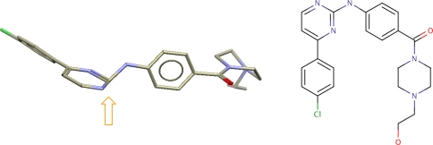
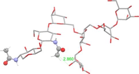
Unusual aminopyrimidine geometry in the ligand in 3qad
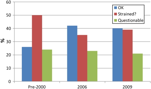
Classification of ligands according to structural geometry for samples of protein/ligand structures submitted to the PDB at different times
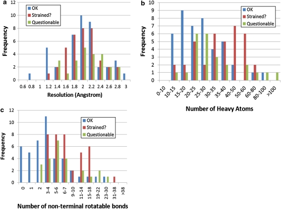
Breakdown of each class a according to resolution of the protein structure; b according to the number of heavy atoms in the ligand, c according to the number of non-terminal rotatable bonds in the ligand
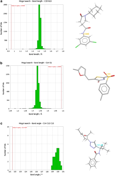
Severe deviations from normal bond and angle geometry: a extremely short bond in 2h7 m ligand; b extremely long S=O bonds in 2hwh ligand; c Very tight C=C–C bond angle in 2hxz ligand
Incorrect cis amide bond geometries (highlighted) in an otherwise high quality ligand structure (2cl2)
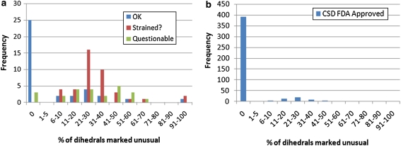
a Breakdown of each ligand class according to percentage of unusual dihedrals. b Breakdown for a set of 440 CSD structures of FDA approved molecules
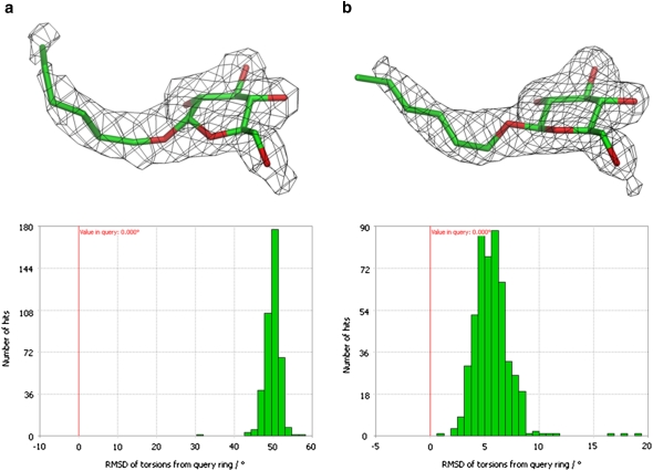
Although the ligand in 2evs (2.2 Å Resolution) apparently fits the e.d. the ring geometry is poor (a). Re-refinement leads to similar quality structure with much improved ring geometry (b)
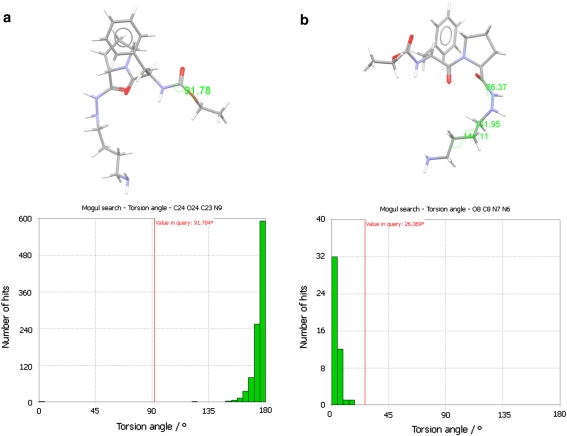
Thrombin inhibitor structure 1ae8 a analysis of the carbamate geometry, b analysis of O=C–N–N (O8 C8 N7 N6) dihedral. This and two other dihedral angles of unusual geometry are highlighted on the 1ae8 ligand structure
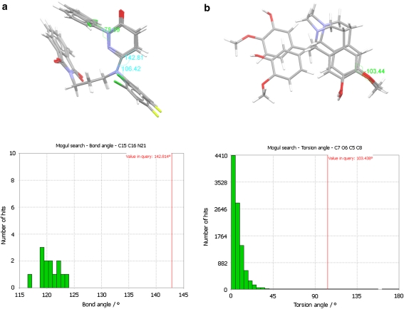
Cases where there is double occupancy: a The CSD distribution of the C-Car-Nar bond angle (C15 C16 N21) in the 2i0 h ligand modelled for double occupancy. This, at 142.8º, is extremely distorted from normal geometry. Other unusual geometric features are labelled in the left-hand image; b the CSD distribution of the CC-OMe (C7 O6 C5 C8) dihedral highlighted on the ligand from 3fwa. This, at 103.44º, is significantly distorted from normal in both ligand placements
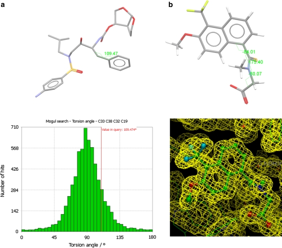
High resolution structures: a The CSD distribution for a typical non-optimum dihedral (C33 C38 C32 C19) in the ligand from the ultra-high resolution structure 2hs1; b the electron density fit and molecular structure for tolrestat in 1zua. The three dihedrals highlighted have chemistry which is matched by only one CSD entry, CAKWAM. However the geometrical correspondence is exact between 1zua and CAKWAM for these three dihedrals
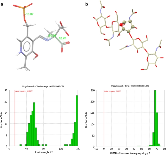
Structures of substrates or analogues: a The NAD-methylaspartate conjugate from 1ajs. Three dihedrals are highly strained (CSD distribution is shown only for the C–O–P–O dihedral); b Penta-NAG bound in chitinase. The central unit takes up an unusual boat form
References
-
- Davis AM, Teague SJ, Kleywegt GJ (2003) Applications and limitations of X-ray crystallographic data in structure-based ligand and drug design. Angew Chem Int Ed 42:2718–2736 - PubMed
MeSH terms
Substances
LinkOut - more resources
Full Text Sources
Research Materials
