. 2012 Apr;27(4):1041-8.
doi: 10.3892/or.2012.1630. Epub 2012 Jan 11.

Carnosic acid inhibits the proliferation and migration capacity of human colorectal cancer cells

Affiliations

Carnosic acid inhibits the proliferation and migration capacity of human colorectal cancer cells

M V Barni et al. Oncol Rep. 2012 Apr.

Abstract

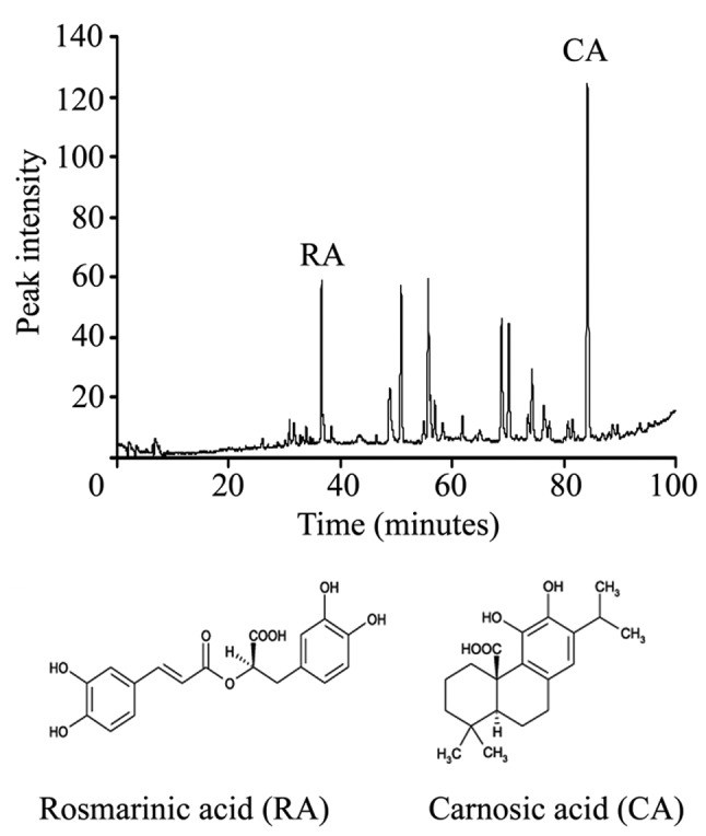
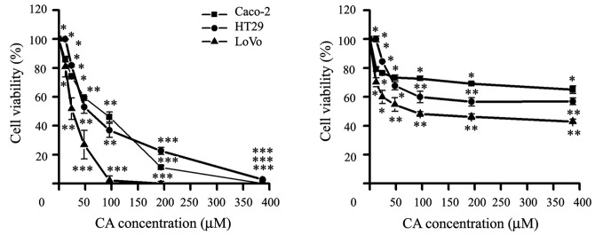
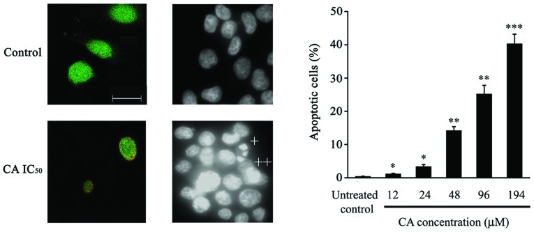
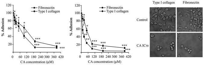
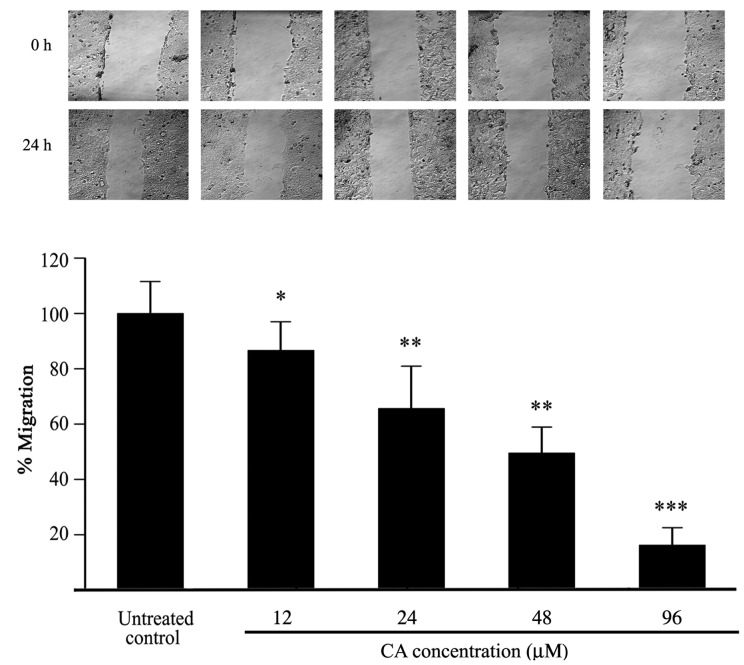
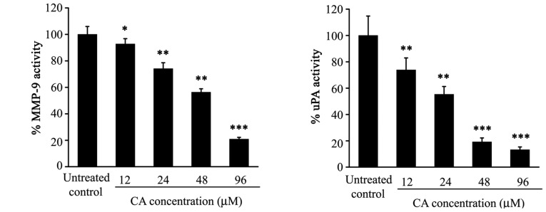
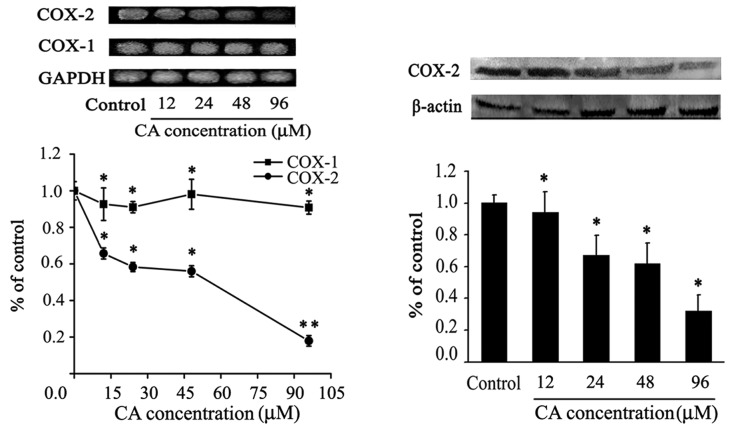
Colorectal cancer (CRC) is the third most common malignant neoplasm worldwide. The objective of this study was to examine whether carnosic acid (CA), the main antioxidant compound of Rosmarinus officinalis L., would inhibit the cell viability of three CRC cell lines: Caco-2, HT29 and LoVo in a dose-dependent manner, with IC₅₀ values in the range of 24-96 µM. CA induced cell death by apoptosis in Caco-2 line after 24 h of treatment and inhibited cell adhesion and migration, possibly by reducing the activity of secreted proteases such as urokinase plasminogen activator (uPA) and metalloproteinases (MMPs). These effects may be associated through a mechanism involving the inhibition of the COX-2 pathway, because we have determined that CA downregulates the expression of COX-2 in Caco-2 cells at both the mRNA and protein levels. Therefore, CA modulates different targets involved in the development of CRC. These findings indicate that carnosic acid may have anticancer activity and may be useful as a novel chemotherapeutic agent.


Figures

Figure 1
Figure 1
Chromatographic profile of the extract of R. officinalis L. determined by HPLC (A). Molecular structures of RA (left) and CA (right) (B).
Figure 2
Figure 2
Effect of CA (A), RA (B) on cell viability of Caco-2, HT29 and LoVo cell lines. Cells were exposed to CA and RA (0 at 388 μM) for 24 h and cell viability was measure by MTS assay. The values represent means ± SD of three cultures from triplicate-independent experiments. (*P<0.05, **P<0.01, ***P<0.001 vs. control).
Figure 3
Figure 3
Effect of CA on Caco-2 apoptotic cell death. (A) CA induces phosphatidylserine translocation, one of the early apoptotic features, in Caco-2 cells. Cells were cultured in absence (control) and presence (CA IC50) for 24 h and exposed to double-stained with 6-CFDA and Annexin-V-Cy3 and photographed with confocal and fluorescence microscopy. Similar results were obtained from two additional separated experiments. Fluorescence microscopic images of Caco-2 cells treated with CA at IC50 for 24 h and stained DAPI dye. (B) CA induces signs of late apoptotic features such as apoptotic bodies (+) and condensation of the nuclear material (++). Scale bar, 20 μm. (C) The percentage of apoptotic cells, stained as indicated in B, was recorded. The values represent mean ± SD of three independents experiments. (*P<0.05, **P<0.01, ***P<0.001 vs. control).
Figure 4
Figure 4
Inhibition of adhesion cells by CA treatment. (A) Caco-2 cells were cultured in 96-well multiplates, previously coated with type I collagen or fibronectin, in the presence or absence of CA (0–388 μM) for 1 h. Then, cells were stained with 2% crystal violet and the optical density at 595 nm was measured. (B) Cells were pre-incubated with CA for 24 h, washed and cultured as described above. (C) Fluorescence-microscope images of untreated and pre-treated cells with CA at IC50 for 24 h were taken after 1 h of incubation onto the coated wells with type I collagen (left) and fibronectin (right). The values represent means ± SD of six cultures from duplicate experiments. (*P<0.05, **P<0.01, ***P<0.001 vs. control). Scale bar, 20 μm.
Figure 5
Figure 5
Effect of CA on the migration of Caco-2 cells. (A) Confluent cell monolayers were wounded with a pipet tip and incubated without or with CA (0 at 388 μM) for 24 h. Wound were monitored and photographed at 0 and 24 h of incubation with a phase-contrast microscope. The percentage of migration is shown in B. The values represent mean ± SD of quadruplicate cultures from duplicate experiments. (*P<0.05, **P<0.01, ***P<0.001 vs. control).
Figure 6
Figure 6
Effect of CA on extracellular matrix components activity of Caco-2 cells. Cells were incubated without or with CA (0 at 96 μM) for 48 h. Samples of conditioned medium were seeded in agarose gel with casein and plasminogen and then were incubated in a humidified chamber at 37°C for 48 h. uPA activities were quantified by radial caseinolisis and determined the percentage of inhibition of uPA activity. The values represent mean ± SD of three experiments (**P<0.01, ***P<0.001 vs. control).
Figure 7
Figure 7
Effect of CA on the expression of COX-2 in Caco-2 cells. (A) Determination of COX-2, COX-1 and GAPDH mRNA by use of RT-PCR. The cells were incubated without or with CA (0 at 96 μM) for 24 h, and subsequently RNA was isolated and reverse transcribed; cDNA of COX-2, COX-1 and GAPDH were amplified by specific primers. The amplified cDNA products were quantified and the percentage of inhibition was determined. (B) The expression of protein COX-2 was assessed by western blot analysis. Levels of COX-2 (top) and β-actin (bottom) were quantified and the percentage of inhibition was determined. The values represent means ± SD of three experiments. (*P<0.05, **P<0.01 vs. control).

Comment in

References

    1. Bruce WR, Giacca A, Medline A. Possible mechanisms relating diet and risk of colon cancer. Cancer Epidemiol Biomarkers Prev. 2000;9:1271–1279. - PubMed
    1. Storz P. Reactive oxygen species in tumor progression. Front Biosci. 2005;10:1881–1896. - PubMed
    1. Kundu JK, Surh YJ. Epigallocatechin gallate inhibits phorbol ester-induced activation of NF-κB and CREB in mouse skin. Ann NY Acad Sci. 2007;1095:504–512. - PubMed
    1. Kellof GJ, Crowell JA, Steele VE, Lubert RA, Malone WA, Boone CW, Koplovich L, Hawk ET, Liberman R, Lawrence JA, Ali I, Viner JL, Sigman CC. Progress in cancer chemoprevention: development of diet-derived chemopreventive agents. J Nutr. 2000;130:S467–S471. - PubMed
    1. Friedl P, Wolf K. Tumor-cell invasion and migration: diversity and escape mechanisms. Nat Rev Cancer. 2003;3:362–374. - PubMed

Publication types

MeSH terms