. 2012 Oct;68(4):1097-108.
doi: 10.1002/mrm.23314. Epub 2012 Jan 13.

Volumetric navigators for real-time motion correction in diffusion tensor imaging

Affiliations

Volumetric navigators for real-time motion correction in diffusion tensor imaging

A Alhamud et al. Magn Reson Med. 2012 Oct.

Abstract

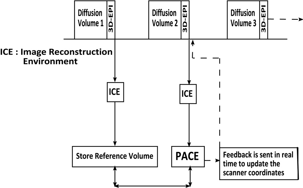
Prospective motion correction methods using an optical system, diffusion-weighted prospective acquisition correction, or a free induction decay navigator have recently been applied to correct for motion in diffusion tensor imaging. These methods have some limitations and drawbacks. This article describes a novel technique using a three-dimensional-echo planar imaging navigator, of which the contrast is independent of the b-value, to perform prospective motion correction in diffusion weighted images, without having to reacquire volumes during which motion occurred, unless motion exceeded some preset thresholds. Water phantom and human brain data were acquired using the standard and navigated diffusion sequences, and the mean and whole brain histogram of the fractional anisotropy and mean diffusivity were analyzed. Our results show that adding the navigator does not influence the diffusion sequence. With head motion, the whole brain histogram-fractional anisotropy shows a shift toward lower anisotropy with a significant decrease in both the mean fractional anisotropy and the fractional anisotropy histogram peak location (P<0.01), whereas the whole brain histogram-mean diffusivity shows a shift toward higher diffusivity with a significant increase in the mean diffusivity (P<0.01), even after retrospective motion correction. These changes in the mean and the shape of the histograms are recovered substantially in the prospective motion corrected data acquired using the navigated sequence.


Figures

Figure 1
Figure 1
Flowchart of the modified diffusion pulse sequence with interleaved 3D-EPI navigator. Data is transferred to ICE, registration is performed by PACE, and the position and gradient system is updated before acquisition of the next diffusion volume.
Figure 2
Figure 2
(A) The MD map for slice 38 of a stationary water phantom scanned using the basic diffusion sequence, and the difference between the MD map of this slice and the MD map of the same slice for scan (B) W_basic2, (C) W_basic3, (D) W_vNav1, (E) W_vNav2, and (F) W_vNav3, where W denotes water phantom scans, basic denotes scans acquired using the basic diffusion sequence, and vNav denotes scans acquired using the navigated prospective motion corrected diffusion sequence. All color bars have units 10−3mm2s−1.
Figure 3
Figure 3
Histograms of the averaged MD for the three scans over the whole volume of the water phantom for the navigated prospective motion corrected diffusion sequence (vNav) and the basic diffusion sequence (basic).
Figure 4
Figure 4
Normalized whole brain histograms (WBHs) of FA for two subjects (2 and 5) for the at rest (NoMo) scans acquired with the basic diffusion sequence and with the navigated sequence without prospective motion correction (vNav_NoCo). The plots in the bottom row are the corresponding motion parameters that were estimated in ICE by PACE during the NoMo_vNav_NoCo scans.
Figure 5
Figure 5
Comparison of motion parameter estimates generated by PACE and retrospective (retro) motion correction for the first subject. Figures 5A and 5D show the motion parameters that were estimated in ICE by PACE for the Mo_vNav_NoCo and Mo_vNav_all scans, respectively. Figures 5B and 5E show the retrospective motion estimates for the Mo_vNav_NoCo scan using SPM and FLIRT, respectively, and same for the basic diffusion sequence in figures 5C and 5F. Mo denotes a scan with motion, vNav the navigated diffusion sequence, NoCo without prospective motion correction, and all a scan with prospective motion correction and reacquisition enabled. Subjects moved upon verbal instruction, 5 to 6 times during the scan.
Figure 6
Figure 6
FA and MD maps of slice 80 for the first subject for 5 different acquisitions, as well as results of retrospective motion correction in the scan acquired using the basic sequence using SPM (Mo_basic_retro_SPM) and FLIRT (Mo_basic_retro_FLIRT), respectively. Data acquired in the Mo_vNav_all scan have been analysed both without (Mo_vNav_noReAq) and with (Mo_vNav_ReAq) reacquisition. The two yellow circles on the FA maps demonstrate reduced blurring in the scan with reacquisition compared to the scan without reacquisition. All the maps are co-registered to the T1 space.
Figure 7
Figure 7
Comparison of the normalized WBH-FA (A–D) and WBH-MD (E–H) for the first subject for different scans. (A,E) comparison of at rest scans acquired using both the basic and navigated sequence to scans with motion and no prospective motion correction acquired using both the basic and navigated sequence; (B,F) effect of retrospective motion correction using SPM on the scans acquired without prospective motion correction; (C,G) effect of retrospective motion correction using FLIRT on the scans acquired without prospective motion correction; and (D,H) prospective motion corrected scans acquired using the navigated sequence, both without and with reacquisition.
Figure 8
Figure 8
Comparison of the normalized WBH-FA in three subjects for an at rest baseline scan (NoMo_basic) compared to a scan with motion and retrospective motion correction where the suffices BE and AE, respectively, denote before and after elimination of corrupted volumes that have low signal due to motion.
Figure 9
Figure 9
(A) The effect of motion on the normalized WBH-FA of the basic diffusion sequence (both before and after retrospective motion correction with FLIRT and SPM) for a particularly restless subject, (B) normalized WBH-FA of the navigated sequence with prospective motion correction, both without and with reacquisition, for this data with many uncorrected corrupted volumes, (C) normalized WBH-FA of the navigated sequence after elimination (AE) of uncorrected corrupted volumes.

Similar articles

Cited by

References

    1. Basser PJ, Pierpaoli C. Microstructural and physiological features of tissues elucidated by quantitative-diffusion-tensor MRI. J Magn Reson B. 1996;111(3):209–219. - PubMed
    1. Hasan KM, Iftikhar A, Kamali A, Kramer LA, Ashtari M, Cirino PT, Papanicolaou AC, Fletcher JM, Ewing-Cobbs L. Development and aging of the healthy human brain uncinate fasciculus across the lifespan using diffusion tensor tractography. Brain Research. 2009;1276:67–76. - PMC - PubMed
    1. Snook L, Paulson LA, Roy D, Phillips L, Beaulieu C. Diffusion tensor imaging of neurodevelopment in children and young adults. NeuroImage. 2005;26(4):1164–1173. - PubMed
    1. Chung S, Pelletier D, Sdika M, Lu Y, Berman JI, Henry RG. Whole brain voxel-wise analysis of single-subject serial DTI by permutation testing. NeuroImage. 2008;39(4):1693–1705. - PMC - PubMed
    1. Schwartzman A, Dougherty RF, Taylor JE. Cross-subject comparison of principal diffusion direction maps. Magn Reson Med. 2005;53(6):1423–1431. - PMC - PubMed

Publication types

MeSH terms