. 2012 Jan 13;335(6065):225-8.
doi: 10.1126/science.1214400.

Tumor necrosis factor signaling requires iRhom2 to promote trafficking and activation of TACE

Affiliations

Tumor necrosis factor signaling requires iRhom2 to promote trafficking and activation of TACE

Colin Adrain et al. Science. .

Abstract

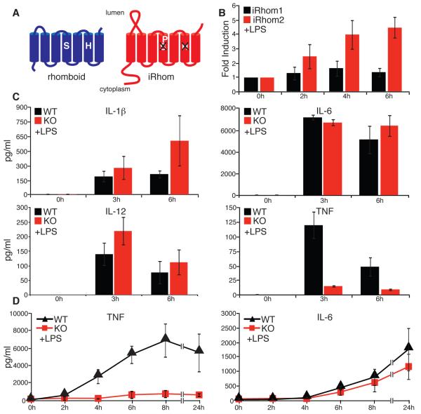
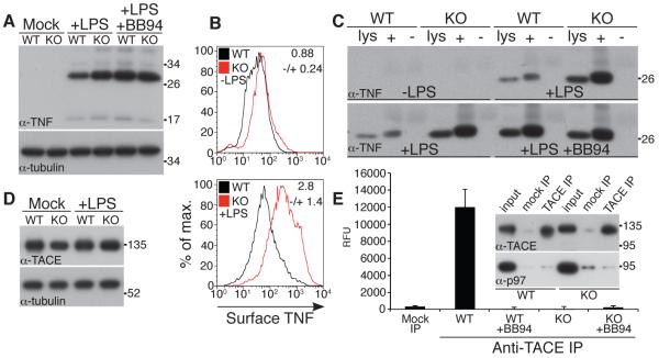
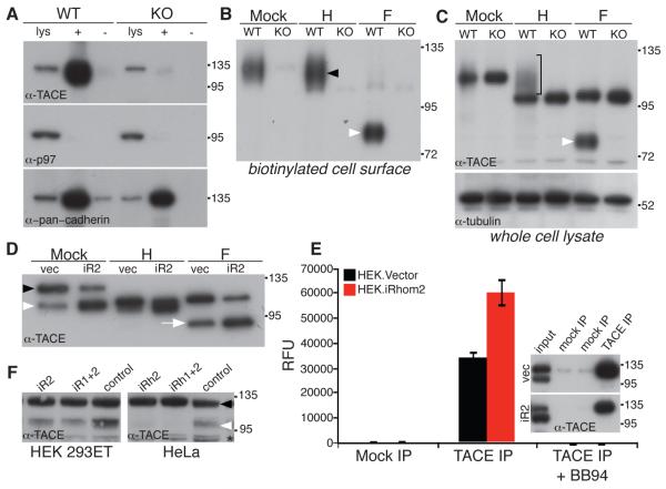
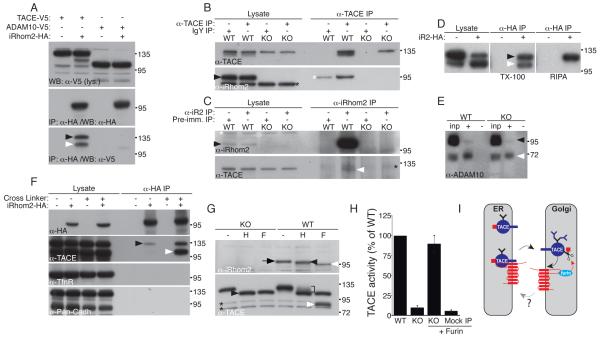
The cytokine tumor necrosis factor (TNF) is the primary trigger of inflammation. Like many extracellular signaling proteins, TNF is synthesized as a transmembrane protein; the active signal is its ectodomain, which is shed from cells after cleavage by an ADAM family metalloprotease, ADAM17 (TNFα-converting enzyme, TACE). We report that iRhom2 (RHBDF2), a proteolytically inactive member of the rhomboid family, is required for TNF release in mice. iRhom2 binds TACE and promotes its exit from the endoplasmic reticulum. The failure of TACE to exit the endoplasmic reticulum in the absence of iRhom2 prevents the furin-mediated maturation and trafficking of TACE to the cell surface, the site of TNF cleavage. Given the role of TNF in autoimmune and inflammatory diseases, iRhom2 may represent an attractive therapeutic target.


Figures

Fig. 1
Fig. 1
iRhom2 is essential for LPS-induced TNF shedding. (A) Comparison of an active rhomboid protease and iRhom. (B) Fold induction of iRhom2 mRNA in response to LPS in WT macrophages (mean of 3 experiments ±SD), as measured by qPCR. mRNA levels were normalized to endogenous actin. (C) Serum concentrations of the indicated cytokines in WT and iRhom2−/− mice (KO) in response to LPS (mean of 5 experiments ±SD), as measured by multi-analyte cytokine profiling. (D) TNF and IL-6 production by LPS-stimulated BMDMs (WT and iRhom2−/− (KO); mean of 3 experiments ±SD), as measured by ELISA. All graphs show mean±SD.
Fig. 2
Fig. 2
TNF biogenesis is normal but TACE proteolytic activity is defective in iRhom2−/− cells. (A) Immunoblots for TNF of extracts from mock, LPS, or LPS/BB94-treated macrophages. Anti-tubulin was used to determine equal loading throughout. (B) Flow cytometric analysis of surface TNF on CD11b+ WT and iRhom2−/− (KO) macrophages 3 hours after LPS treatment. The fold-change in mean fluorescent intensity (KO/WT) is indicated in each graph (mean of 3 experiments ±SD). (C) Whole cell lysate (lys) versus cell surface biotinylated (+) or mock-treated (−) proteins bound to neutravidin resin from WT or KO macrophages were immunoblotted as indicated. (D) Immunoblot of TACE expression in BMDMs. (E) Immunoprecipitates from iRhom2−/− macrophages subjected to a fluorogenic cleavage assay (mean of 3 experiments ±SD). Specificity of the TACE antibody is demonstrated by probing IPs with TACE and p97 antibodies (inset in E). Levels of TACE in the activity assay were equal in WT and KO macrophages (inset E and Fig. S2B).
Fig. 3
Fig. 3
iRhom2 is essential for ER exit and activation of TACE. (A) Cell surface biotinylation was used to detect TACE at the plasma membrane in WT and iRhom2−/− (KO) macrophages: whole cell lysates (lys); neutravidin captured cell surface proteins (+); mock-treated (−). Cytoplasmic protein p97 was used as a negative control for cell surface biotinylation; surface levels of cadherin are comparable in WT and KO cells. (B) Biotinylated cell surface proteins were deglycosylated with endoglycosidase-H (H), which cleaves ER but not Golgi N-glycans, or PNGase F (F) which removes both. Cell surface TACE is endo-H resistant (black arrow). TACE matures by acquiring Golgi N-glycans, and furin-catalyzed removal of the prodomain. PNGase F treatment of cell surface TACE generates a lower molecular weight polypeptide corresponding to the furin-cleaved species (white arrowhead; see also Fig. S2B,C). (C) TACE immunoblot of whole cells lysates treated with endo-H (H) or PNGase F (F) Bracket: endo-H resistant mature TACE. For PNGase F-treated lysates: the upper band is ER resident, immature TACE, and the lower band is furin-cleaved TACE (white arrow, see also Fig. 3B, S2C,D). (D) Cell extracts from HEK cells stably expressing mouse iRhom2 were deglycosylated with endo-H (H) or PNGase F (F) and immunoblotted for TACE. Unlike mouse BMDM-derived TACE, human TACE can be detected as two species in lysates without prior deglycosylation (immature, black arrowhead; mature, white arrowhead). (E) Activity of TACE immunoprecipitates from HEK cells expressing iRhom2 (mean of 3 experiments ±SD); specificity of the IP was confirmed by western blot (inset). (F) The effect of siRNA knockdown of human iRhom2 (and combined iRhom1/2) on the expression of endogenous mature TACE (white arrowhead; black arrowhead indicates immature TACE) in human HEK 293T and HeLa cells. As a control, an unrelated member of the rhomboid family (RHBDL2) was knocked down. The asterisk denotes a non-specific band.
Fig. 4
Fig. 4
iRhom2 interacts with TACE and leaves the ER. (A) Immunoprecipitates from HEK cells or HEK cells stably expressing iRhom2-HA, transfected with TACE-V5 or ADAM10-V5, were immunoblotted for iRhom2. Co-immunoprecipitation of immature and mature TACE is indicated by black and white arrowheads, respectively. (B) Macrophage lysates were immunoprecipitated with anti-TACE and immunoblotted for iRhom2 or TACE. Arrowhead: endogenous iRhom2. Black asterisk: a non-specific band detected in both KO and WT. White asterisk: background binding of iRhom2 to the resin. (C) Macrophage lysates were immunoprecipitated with anti-iRhom2 and immunoblotted for iRhom2 or TACE. White arrowhead: endogenous TACE. Black arrowhead: endogenous iRhom. White asterisk: original TACE signal detected following reprobing. Black asterisk: a non-specific band in all IP lanes. (D) HEK cells expressing iRhom2-HA were immunoprecipitated with anti-HA and immunoblotted for TACE. IPs were performed under mild (Triton X-100 detergent), and more stringent (RIPA buffer) conditions. (E) Complete input extracts (inp) and surface biotinylated proteins (+) from WT and iRhom2 KO macrophages immunoblotted for ADAM10. Black arrow: immature ADAM10; white arrow: mature ADAM10. (F) Immunoprecipitates (in RIPA buffer) of iRhom2-HA from HEK cells; precipitates were done +/− the crosslinker DSP. Transferrin receptor and pan-cadherin immunoblots were used as negative controls. (G) Lysates from LPS-treated WT or KO macrophages were treated with endo-H (H) or PNGase-F (F), and immunoblotted for iRhom2. Arrow: fully glycosylated iRhom2. Black and white arrowheads: endo-H-resistant single glycan form and unmodified polypeptide, respectively (iRhom2 has two predicted N-linked glycosylation sites; only one is modified in the Golgi). Lower panel: TACE in the same lysates as control. Black arrowhead: endo-H-sensitive TACE. Bracket: endo-H-resistant TACE. White arrowhead: mature TACE. Asterisks: non-specific bands. (H) TACE immunoprecipitates were preincubated −/+ 100 nM recombinant furin prior to measuring TACE activity. (I) Model of iRhom2 function in macrophages.

Comment in

  • Cell biology. Sheddase gets guidance.
    Lichtenthaler SF. Lichtenthaler SF. Science. 2012 Jan 13;335(6065):179-80. doi: 10.1126/science.1216815. Science. 2012. PMID: 22246765 No abstract available.

References

    1. Gooz M. Crit. Rev. Biochem. Mol. Biol. 2010;45:146. - PMC - PubMed
    1. Peschon JJ, et al. Science. 1998;282:1281. - PubMed
    1. Tracey D, Klareskog L, Sasso EH, Salfeld JG, Tak PP. Pharmacol. Ther. 2008;117:244. - PubMed
    1. Black RA, et al. Nature. 1997;385:729. - PubMed
    1. Le Gall SM, Maretzky T, Issuree PD, Niu XD, Reiss K, Saftig P, Khokha R, Lundell D, Blobel CP. J. Cell Sci. 2010;123:3913. - PMC - PubMed

Publication types