Review
. 2012 May;35(5):1026-37.
doi: 10.1002/jmri.23581. Epub 2012 Jan 13.

Applications of arterial spin labeled MRI in the brain

Affiliations
Review

Applications of arterial spin labeled MRI in the brain

John A Detre et al. J Magn Reson Imaging. 2012 May.

Abstract

Perfusion provides oxygen and nutrients to tissues and is closely tied to tissue function while disorders of perfusion are major sources of medical morbidity and mortality. It has been almost two decades since the use of arterial spin labeling (ASL) for noninvasive perfusion imaging was first reported. While initial ASL magnetic resonance imaging (MRI) studies focused primarily on technological development and validation, a number of robust ASL implementations have emerged, and ASL MRI is now also available commercially on several platforms. As a result, basic science and clinical applications of ASL MRI have begun to proliferate. Although ASL MRI can be carried out in any organ, most studies to date have focused on the brain. This review covers selected research and clinical applications of ASL MRI in the brain to illustrate its potential in both neuroscience research and clinical care.


Figures

Figure 1
Figure 1
Typical whole-brain ASL MRI quantitative CBF data obtained in 6 minutes at 3 Tesla using PASL and pCASL with echoplanar imaging (TOP and MIDDLE), adapted from (109) with permission from the publisher. BELOW: pCASL with background-suppressed 3-dimensional variable density spiral acquisition acquired in 2 minutes at 3 Tesla.
Figure 2
Figure 2
Transit artifact in a patient with left middle cerebral artery stroke and a transit time map showing prolonged arterial transit to this region. TOP: FLAIR images showing multiple strokes in the left MCA distribution. MIDDLE: ASL CBF images show artifactual hyperperfusion in the left MCA distribution (arrows) due to delayed transit of label, which is imaged within leptomenigeal vessels providing collateral flow. CBF in left and right MCA distributions are actually nearly identical at 43 and 42 ml/100g/min, respectively. Bottom: Arterial transit time map demonstrates prolonged transit times to the left MCA distribution.
Figure 3
Figure 3
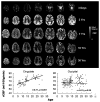
TOP: Representative ASL MRI data across human brain development. BELOW: ROI data showing developmental trajectories of relative CBF in cingulate and occipital cortices. An increase in cingulate CBF is evident, while occipital CBF remains stable.
Figure 4
Figure 4
Temporal stability of ASL perfusion fMRI. Successful demonstration of motor cortex activation with bilateral finger tapping is observed even when task and activation are carried out on successive days, 24 hours apart. The experimental design is shown above. Adapted from (84) with permission from the publisher.
Figure 5
Figure 5
Demonstration of genotype and phenotype effects in resting ASL MRI data. LEFT: increased perfusion of amygdala in patients with a serotonin transporter genotype that confers an increased risk of depression and anxiety. RIGHT: Resting perfusion in right medial frontal cortex predicts subsequent time-on-task fatiguability. Adapted from (88,94) with permission from the publisher.
Figure 6
Figure 6
Increase in orbitofrontal cortex CBF after overnight abstinence in smokers (LEFT) and reduction in CBF in this region after treatment with baclofen (RIGHT). Adapted from (121,129) with permission from the publisher.

References

    1. Ackerman JJ, Ewy CS, Becker NN, Shalwitz RA. Deuterium nuclear magnetic resonance measurements of blood flow and tissue perfusion employing 2H2O as a freely diffusible tracer. Proc Natl Acad Sci U S A. 1987;84(12):4099–4102. - PMC - PubMed
    1. Detre JA, Subramanian VH, Mitchell MD, et al. Measurement of regional cerebral blood flow in cat brain using intracarotid 2H2O and 2H NMR imaging. Magn Reson Med. 1990;14(2):389–395. - PubMed
    1. Eleff SM, Schnall MD, Ligetti L, et al. Concurrent measurement of cerebral blood flow, sodium, lactate, and high-energy phosphate metabolism using 19F, 23Na, 1H, and 31P nuclear magnetic resonance spectroscopy. Magn Reson Med. 1988;7(4):412–424. - PubMed
    1. Detre JA, Eskey CJ, Koretsky AP. Measurement of cerebral blood flow in rat brain using trifluoromethane and 19F-NMR. Magn Reson Med. 1990;15(1):45–57. - PubMed
    1. Detre JA, Leigh JS, Williams DS, Koretsky AP. Perfusion imaging. Magn Reson Med. 1992;23(1):37–45. - PubMed

Publication types