. 2012 May;46(5):687-94.
doi: 10.1165/rcmb.2011-0040OC. Epub 2012 Jan 12.

Spiruchostatin A inhibits proliferation and differentiation of fibroblasts from patients with pulmonary fibrosis

Affiliations

Spiruchostatin A inhibits proliferation and differentiation of fibroblasts from patients with pulmonary fibrosis

Elizabeth R Davies et al. Am J Respir Cell Mol Biol. 2012 May.

Abstract

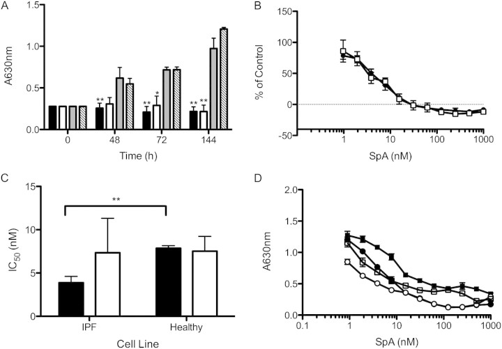
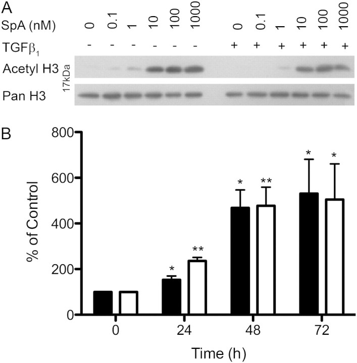
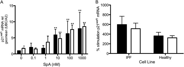
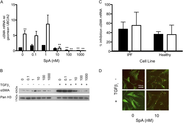
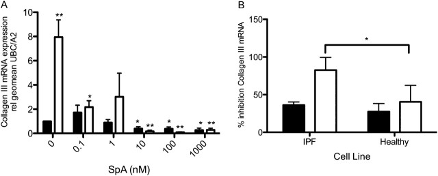
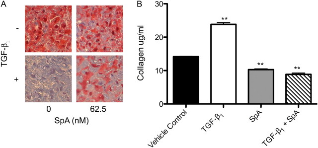
Idiopathic pulmonary fibrosis (IPF) is a progressive scarring disorder characterized by the proliferation of interstitial fibroblasts and the deposition of extracellular matrix causing impaired gas exchange. Spiruchostatin A (SpA) is a histone deacetylase inhibitor (HDI) with selectivity toward Class I enzymes, which distinguishes it from other nonspecific HDIs that are reported to inhibit (myo)fibroblast proliferation and differentiation. Because the selectivity of HDIs may be important clinically, we postulated that SpA inhibits the proliferation and differentiation of IPF fibroblasts. Primary fibroblasts were grown from lung biopsy explants obtained from patients with IPF or from normal control subjects, using two-dimensional or three-dimensional culture models. The effect of SpA on fibroproliferation in serum-containing medium ± transforming growth factor (TGF)-β(1) was quantified by methylene blue binding. The acetylation of histone H3, the expression of the cell-cycle inhibitor p21(waf1), and the myofibroblast markers α-smooth muscle actin (α-SMA) and collagens I and III were determined by Western blotting, quantitative RT-PCR, immunofluorescent staining, or colorimetry. SpA inhibited the proliferation of IPF or normal fibroblasts in a time-dependent and concentration-dependent manner (concentration required to achieve 50% inhibition = 3.8 ± 0.4 nM versus 7.8 ± 0.2 nM, respectively; P < 0.05), with little cytotoxicity. Western blot analyses revealed that SpA caused a concentration-dependent increase in histone H3 acetylation, paralleling its antiproliferative effect. SpA also increased p21(waf1) expression, suggesting that direct cell-cycle regulation was the mechanism of inhibiting proliferation. Although treatment with TGF-β(1) induced myofibroblast differentiation associated with increased expression of α-SMA, collagen I and collagen III and soluble collagen release, these responses were potently inhibited by SpA. These data support the concept that bicyclic tetrapeptide HDIs merit further investigation as potential treatments for IPF.


Figures

Figure 1.
Figure 1.
Inhibition of fibroblast proliferation by spiruchostatin A (SpA). Idiopathic pulmonary fibrosis (IPF) fibroblasts were cultured for up to 144 hours in DMEM/FBS and SpA. (A) Time-course data for fibroblasts from a single IPF donor, assessing the antiproliferative effect of 10 nM SpA in the absence (solid bar) or presence (open bar) of transforming growth factor (TGF)–β1 compared with vehicle control alone (grey bar) or vehicle in the presence of TGF-β1 (hatched bar). (B) Growth inhibition plots for cells from the same donor, grown in the absence (solid circles) or presence (open squares) of TGF-β1 for 144 hours and treated with a range of SpA concentrations. The cell number was expressed as a percentage of the vehicle control at 144 hours, after subtraction of the cell number at t = 0. (C) The concentrations required to achieve 50% inhibition (IC50 values) for the antiproliferative effects of SpA, determined from dose–response curves for fibroblasts from three IPF donors and three normal control subjects (as shown in Figure E2). (D) The persistence of the effect of SpA (48 hours of exposure) on fibroblast proliferation in the absence (solid circles) or presence (open squares) of TGF-β1, measured 96 hours after the removal of SpA. For comparison, fibroblasts were continuously exposed to SpA in the absence (solid squares) or presence (open circles) of TGF-β1. Data are expressed as in B. Data represent means ± SD (n = 3 individual experiments). *P < 0.05, versus corresponding control sample. **P < 0.01, versus corresponding control sample (i.e., vehicle without or with TGF-β1).
Figure 2.
Figure 2.
SpA increases histone H3 acetylation in primary fibroblasts. IPF fibroblasts were cultured in DMEM/FBS ± TGF-β1, with the indicated concentrations of SpA at 24, 48, or 72 hours before harvesting. Cells were analyzed by Western blotting, using a primary antibody against acetyl histone H3, with a pan histone H3 antibody as loading control. (A) Western blots obtained after 48 hours of treatment with different doses of SpA. (B) Semiquantitative analysis of the time-course analysis of histone acetylation in the presence of 10 nM SpA and in the absence (solid bar) or presence (open bar) of TGF-β1, determined using densitometry. The data represent means ± SD (n = 3 separate experiments). *P < 0.05, versus corresponding control sample. **P < 0.01, versus corresponding control sample, denoting an increase in histone acetylation.
Figure 3.
Figure 3.
SpA increases the fibroblast expression of the cell-cycle regulator p21waf1. RNA was isolated from IPF (A and B) or normal (B) fibroblasts treated with the indicated concentrations of SpA (A), or with 10 nM SpA (B), without TGF-β1 (solid bars) or with TGF-β1 (open bars) for 48 hours. cDNA was synthesized for quantitative PCR analysis. p21waf1 mRNA expression was calculated relative to the geometric mean of the housekeeping genes UBC/A2, using the ΔΔCT method. The data in A are expressed as fold stimulation relative to the untreated control sample (minus TGF-β1), and represent means ± SD (n = 3 separate experiments), using one IPF fibroblast cell line. The data in B were calculated as percent stimulation by SpA, compared with expression in the absence or presence of TGF-β alone, and are shown as means ± SD from three IPF and three normal fibroblast lines (n = 3 per subject). *P < 0.05, versus corresponding control sample. **P < 0.01, versus corresponding control sample (i.e., vehicle with or without TGF-β).
Figure 4.
Figure 4.
TGF-β1 induces α–smooth muscle actin (α-SMA) mRNA and protein expression, which can be inhibited by SpA. IPF (A–D) or normal (C) fibroblasts were cultured without (solid bars) or with (open bars) TGF-β1, and with the indicated concentrations of SpA (A and B) or 10 nM SpA (C) before harvesting for RNA or protein extraction at the appropriate time point. (A and C) α-SMA mRNA expression was analyzed by quantitative RT-PCR and calculated relative to the geometric mean of the housekeeping genes UBC/A2, using the ΔΔCT method. The data in A are expressed as fold stimulation relative to the untreated control sample, and represent means ± SD (n = 3 separate experiments), using one IPF fibroblast cell line. The data in C were calculated as percent inhibition of gene expression by SpA compared with that seen in the absence or presence of TGF-β1 alone, and are shown as means ± SD from three IPF and three normal fibroblast lines (n = 3 per subject). (B) α-SMA protein expression at 72 hours was analyzed by Western blotting, with histone H3 as a loading control. (D) Immunofluorescent staining of α-SMA in fibroblasts, treated in the absence or presence of TGF-β1 ± SpA (10 nM) for 48 hours, as indicated. Nuclei were counterstained with 7-amino actinomycin D (red). *P < 0.05, versus corresponding control sample. **P < 0.01, versus corresponding control sample.
Figure 5.
Figure 5.
Effects of SpA on interstitial collagen expression. RNA was isolated from IPF fibroblasts treated without (solid bars) or with (open bars) TGF-β1, and with the indicated doses of SpA (A) or 10 nM SpA (B) for 48 hours. Collagen III mRNA expression was measured by quantitative RT-PCR and calculated relative to the housekeeping genes UBC/A2, using the ΔΔCT method. Data in A are expressed as fold stimulation relative to the untreated control sample, and represent means ± SD (n = 3 separate experiments), using one IPF fibroblast cell line. Data in B were calculated as percent inhibition of gene expression by SpA compared with that seen in the absence or presence of TGF-β1 alone, and are shown as means ± SD from three IPF and three normal fibroblast lines (n = 3 per subject). *P < 0.05, versus corresponding control sample. **P < 0.01, versus corresponding control sample.
Figure 6.
Figure 6.
SpA diminishes TGF-β1–induced collagen production. Three-dimensional pellet cultures were treated without or with TGF-β1 and 62.5 nM SpA before fixing, paraffin-embedding, and trichrome staining for collagen expression. (A) Histochemical analysis of pellets cultured under indicated conditions, where the binding of aniline blue in Masson trichrome stain denotes the presence of collagen (magnification, ×40). (B) Release of soluble collagen by cultured fibroblasts under the conditions indicated was assessed at 72 hours, using Sirius red dye. Data represent means ± SD (n = 3 individual experiments). **P < 0.01, versus corresponding control sample.

References

    1. Valeyre D, Freynet O, Dion G, Bouvry D, Annesi-Maesano I, Nunes H. Epidemiology of interstitial lung diseases. Presse Med 2010;39:53–59. - PubMed
    1. Meltzer EB, Noble PW. Idiopathic pulmonary fibrosis. Orphanet J Rare Dis 2008;3:8. - PMC - PubMed
    1. American Thoracic Society, European Respiratory Society. American Thoracic Society/European Respiratory Society international multidisciplinary consensus classification of the idiopathic interstitial pneumonias. Am J Respir Crit Care Med 2002;165:277–304. - PubMed
    1. Raghu G, Weycker D, Edelsberg J, Bradford WZ, Oster G. Incidence and prevalence of idiopathic pulmonary fibrosis. Am J Respir Crit Care Med 2006;174:810–816. - PubMed
    1. King TE, Schwarz MI, Brown K, Tooze JA, Colby TV, Waldron JA, Flint A, Thurlbeck W, Cherniack RM. Idiopathic pulmonary fibrosis: relationship between histopathologic features and mortality. Am J Respir Crit Care Med 2001;164:1025–1032. - PubMed

Publication types

MeSH terms