

Accuracy of MR elastography and anatomic MR imaging features in the diagnosis of severe hepatic fibrosis and cirrhosis
- PMID: 22246952
- PMCID: PMC3495186
- DOI: 10.1002/jmri.23585
Accuracy of MR elastography and anatomic MR imaging features in the diagnosis of severe hepatic fibrosis and cirrhosis
Abstract
Purpose: To compare the diagnostic accuracy of magnetic resonance imaging elastography (MRE) and anatomic MRI features in the diagnosis of severe hepatic fibrosis and cirrhosis.
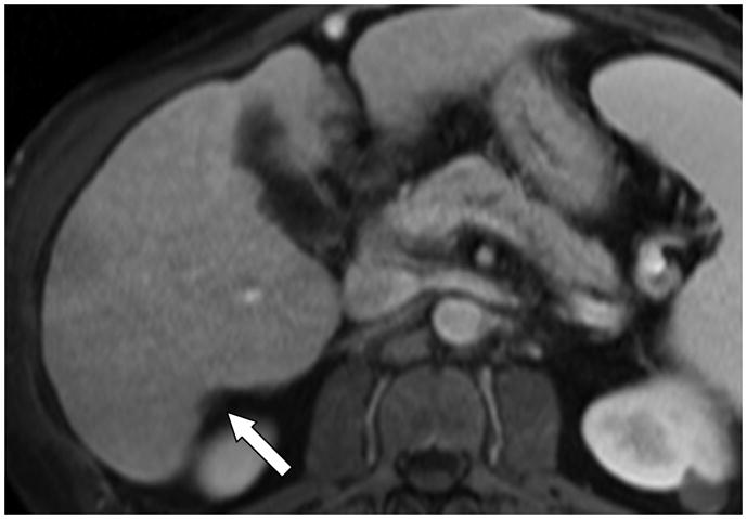
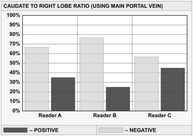
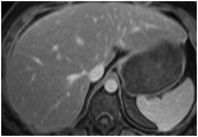
Materials and methods: Three readers independently assessed presence of morphological changes associated with hepatic fibrosis in 72 patients with liver biopsy including: caudate to right lobe ratios, nodularity, portal venous hypertension (PVH) stigmata, posterior hepatic notch, expanded gallbladder fossa, and right hepatic vein caliber. Three readers measured shear stiffness values using quantitative shear stiffness maps (elastograms). Sensitivity, specificity, and diagnostic accuracy of stiffness values and each morphological feature were calculated. Interreader agreement was summarized using weighted kappa statistics. Intraclass correlation coefficient was used to assess interreader reproducibility of stiffness measurements. Binary logistic regression was used to assess interreader variability for dichotomized stiffness values and each morphological feature.
Results: Using 5.9 kPa as a cutoff for differentiating F3-F4 from F0-2 stages, overall sensitivity, specificity, and diagnostic accuracy for MRE were 85.4%, 88.4%, and 87%, respectively. Overall interreader agreement for stiffness values was substantial, with an insignificant difference (P = 0.74) in the frequency of differentiating F3-4 from F0-2 fibrosis. Only hepatic nodularity and PVH stigmata showed moderately high overall accuracy of 69.4% and 72.2%. Interreader agreement was substantial only for PVH stigmata, moderate for C/R m, deep notch, and expanded gallbladder fossa. Only posterior hepatic notch (P = 0.82) showed no significant difference in reader rating.
Conclusion: MRE is a noninvasive, accurate, and reproducible technique compared with conventional features of detecting severe hepatic fibrosis.
Copyright © 2012 Wiley Periodicals, Inc.
Figures


References
-
- Harbin WP, Robert NJ, Ferrucci JT., Jr Diagnosis of cirrhosis based on regional changes in hepatic morphology: a radiological and pathological analysis. Radiology. 1980;135(2):273–83. - PubMed
-
- Giorgio A, Amoroso P, Lettieri G. Cirrhosis: Value of caudate to right lobe ratio in diagnosis with US. Radiology. 1986;161:443–445. - PubMed
-
- Torres WE, Whitmire LF, Gedgaudas-McClees K, et al. Computed tomography of hepatic morphologic changes in cirrhosis of the liver. J Comput Assist Tomogr. 1986;10(1):47–50. - PubMed
-
- Aguirre DA, Behling CA, Alpert E, et al. Liver fibrosis: Noninvasive diagnosis with double contrast material – enhanced MR imaging. Radiology. 2006;239(2):425–437. - PubMed
-
- Fisher MR, Gore RM. Computed tomography in the evaluation of cirrhosis and portal hypertension. J Clin Gastroenterol. 1985;7(2):173–81. - PubMed
Publication types
MeSH terms
Grants and funding
LinkOut - more resources
Full Text Sources
Medical
Miscellaneous
