Review
. 2011 Dec 15;124(Pt 24):4147-59.
doi: 10.1242/jcs.097063.

GLUT4 exocytosis

Affiliations
Review

GLUT4 exocytosis

Jacqueline Stöckli et al. J Cell Sci. .

Abstract

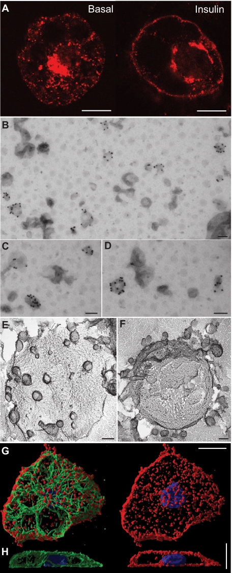
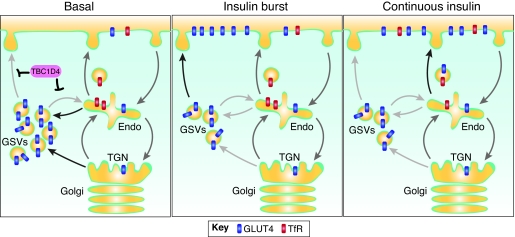
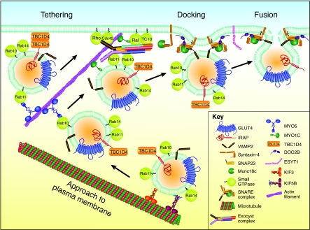
GLUT4 is an insulin-regulated glucose transporter that is responsible for insulin-regulated glucose uptake into fat and muscle cells. In the absence of insulin, GLUT4 is mainly found in intracellular vesicles referred to as GLUT4 storage vesicles (GSVs). Here, we summarise evidence for the existence of these specific vesicles, how they are sequestered inside the cell and how they undergo exocytosis in the presence of insulin. In response to insulin stimulation, GSVs fuse with the plasma membrane in a rapid burst and in the continued presence of insulin GLUT4 molecules are internalised and recycled back to the plasma membrane in vesicles that are distinct from GSVs and probably of endosomal origin. In this Commentary we discuss evidence that this delivery process is tightly regulated and involves numerous molecules. Key components include the actin cytoskeleton, myosin motors, several Rab GTPases, the exocyst, SNARE proteins and SNARE regulators. Each step in this process is carefully orchestrated in a sequential and coupled manner and we are beginning to dissect key nodes within this network that determine vesicle-membrane fusion in response to insulin. This regulatory process clearly involves the Ser/Thr kinase AKT and the exquisite manner in which this single metabolic process is regulated makes it a likely target for lesions that might contribute to metabolic disease.


Figures

Fig. 1.
Fig. 1.
A timeline highlighting some of the key events in research on insulin-regulated glucose transport (Bai et al., 2007; Birnbaum, 1989; Cain et al., 1992; Charron et al., 1989; Cheatham et al., 1996; Cong et al., 1997; Coster et al., 2004; Cushman and Wardzala, 1980; Czech et al., 1993; Dawson et al., 2001; Ebstensen and Plagemann, 1972; Fukumoto et al., 1989; Govers et al., 2004; Hayashi et al., 1998; Huang et al., 2007; James et al., 1988; James et al., 1989; Kaestner et al., 1989; Kanai et al., 1993; Kandror and Pilch, 1994; Karylowski et al., 2004; Katz et al., 1995; Kohn et al., 1998; Kotani et al., 1995; Kurth-Kraczek et al., 1999; Lampson et al., 2001; Lizunov et al., 2005; Lundsgaard, 1939; Martin and Carter, 1970; Martin et al., 1998; Mastick et al., 1994; Merrill et al., 1997; Mueckler et al., 1985; Olson et al., 1997; Quon et al., 1994; Rea et al., 1998; Rodbell, 1964; Sano et al., 2003; Satoh et al., 1993; Shimizu and Shimazu, 1994; Slot et al., 1991a; Slot et al., 1991b; Suzuki and Kono, 1980; Tellam et al., 1997; Volchuk et al., 1996; Wardzala and Jeanrenaud, 1983; Yang et al., 1992). Image of E. Lundsgaard reproduced with permission from BioZoom, published by the Danish Society for Biochemistry and Molecular Biology. Image of ‘Translocation Hypothesis’ was originally published in The Journal of Biological Chemistry (Karnieli et al., 1981) © American Society for Biochemistry and Molecular Biology. Image of GLUT1 is from Mueckler et al. (Mueckler et al., 1985). Reprinted with permission from The American Association for the Advancement of Science.
Fig. 2.
Fig. 2.
Imaging GLUT4 and the cytoskeleton in adipocytes. (A) Immunofluorescence image of untreated (left) or insulin-stimulated (right) 3T3-L1 adipocytes that were immunolabelled for endogenous GLUT4. (BD) Immuno-EM of GLUT4 in vesicles that were prepared from non-stimulated rat adipocytes. After adsorption to EM grids the vesicles were labelled with an antibody against GLUT4, followed by protein-A–gold (10 nm). The size of GLUT4 vesicles in B ranges from 63 to 135 nm. Images were taken by Sally Martin [see Martin et al. (Martin et al., 1997) for methodological details]. (E,F) Deep-etch images of GLUT4 vesicles immunopurified using Staphylococcus aureus cells coated with an anti-GLUT4 antibody then incubated with an adipocyte microsomal fraction. Non-GLUT4-containing vesicles were washed away. Consistent with B–D, the majority of vesicles are small (~50–70 nm) but a population of larger vesicles was also detected. Images were taken by John Heuser using a published method (Rodnick et al., 1992). (G,H) 3D shadow renderings of 120 confocal z-sections (Imaris, Bitplane) of an untreated 3T3-L1 adipocyte. Tubulin (stained with an anti-tubulin antibody) is shown in green, F-actin (stained with TRITC–phalloidin) is shown in red and the nucleus (labelled with DAPI) is shown in blue. Viewed from the top (G) or from the side (H). Scale bars: 10 μm (A,G,H) and 100 nm (B–F).
Fig. 3.
Fig. 3.
A model for GLUT4 trafficking. In the absence of insulin (basal), a pool of GLUT4 is targeted to GSVs, which are derived from the TGN and/or endosomes. In the presence of insulin, these GSVs fuse directly with the plasma membrane in an initial burst (insulin burst). GLUT4 subsequently recycles through endosomes, the TGN and back to the plasma membrane through generic recycling compartments in the presence of insulin (‘continuous insulin’). Following insulin withdrawal, GLUT4 traffics through the TGN to re-form GSVs. Arrows represent different trafficking rates: low (light grey), medium (dark grey), high (black). Endo, endosomes.
Fig. 4.
Fig. 4.
Overview of the steps involved in GLUT4 trafficking at the plasma membrane. GSVs interact with a host of molecules as they are delivered to the cell periphery and tethered, docked and fused at the plasma membrane. GSVs approach the plasma membrane where they are tethered by actin, the exocyst or TBC1D4, or a combination of these in parallel or in series. The vesicle docks with the plasma membrane as the ternary complex is formed between VAMP2 on the GSV and syntaxin-4 and SNAP23 on the plasma membrane. This complex can then facilitate fusion of a GSV with the plasma membrane. The Rab proteins shown on GSVs in this figure were identified on GSVs in at least two separate proteomic studies (see www.jameslab.com.au/Contentpages/DataResources/GSVProteome.shtml).

Similar articles

Cited by

References

    1. Abel E. D., Peroni O., Kim J. K., Kim Y. B., Boss O., Hadro E., Minnemann T., Shulman G. I., Kahn B. B. (2001). Adipose-selective targeting of the GLUT4 gene impairs insulin action in muscle and liver. Nature 409, 729-733 - PubMed
    1. Aledo J. C., Darakhshan F., Hundal H. S. (1995). Rab4, but not the transferrin receptor, is colocalized with GLUT4 in an insulin-sensitive intracellular compartment in rat skeletal muscle. Biochem. Biophys. Res. Commun. 215, 321-328 - PubMed
    1. Antonescu C. N., Diaz M., Femia G., Planas J. V., Klip A. (2008). Clathrin-dependent and independent endocytosis of glucose transporter 4 (GLUT4) in myoblasts: regulation by mitochondrial uncoupling. Traffic 9, 1173-1190 - PubMed
    1. Aran V., Bryant N. J., Gould G. W. (2011). Tyrosine phosphorylation of Munc18c on residue 521 abrogates binding to Syntaxin 4. BMC Biochem. 12, 19 - PMC - PubMed
    1. Babbey C. M., Bacallao R. L., Dunn K. W. (2010). Rab10 associates with primary cilia and the exocyst complex in renal epithelial cells. Am. J. Physiol. Renal Physiol. 299, F495-F506 - PMC - PubMed

Publication types

Substances