. 2012 Jan 31;109(5):1778-83.
doi: 10.1073/pnas.1112500109. Epub 2012 Jan 12.

Isoprenoid biosynthesis is required for miRNA function and affects membrane association of ARGONAUTE 1 in Arabidopsis

Affiliations

Isoprenoid biosynthesis is required for miRNA function and affects membrane association of ARGONAUTE 1 in Arabidopsis

Peter Brodersen et al. Proc Natl Acad Sci U S A. .

Erratum in

Abstract

Plant and metazoan microRNAs (miRNAs) guide ARGONAUTE (AGO) protein complexes to regulate expression of complementary RNAs via base pairing. In the plant Arabidopsis thaliana, the main miRNA effector is AGO1, but few other factors required for miRNA activity are known. Here, we isolate the genes defined by the previously described miRNA action deficient (mad) mutants, mad3 and mad4. Both genes encode enzymes involved in isoprenoid biosynthesis. MAD3 encodes 3-hydroxy-3-methylglutaryl CoA reductase (HMG1), which functions in the initial C(5) building block biogenesis that precedes isoprenoid metabolism. HMG1 is a key regulatory enzyme that controls the amounts of isoprenoid end products. MAD4 encodes sterol C-8 isomerase (HYDRA1) that acts downstream in dedicated sterol biosynthesis. Using yeast complementation assays and in planta application of lovastatin, a competitive inhibitor of HMG1, we show that defects in HMG1 catalytic activity are sufficient to inhibit miRNA activity. Many isoprenoid derivatives are indispensable structural and signaling components, and especially sterols are essential membrane constituents. Accordingly, we provide evidence that AGO1 is a peripheral membrane protein. Moreover, specific hypomorphic mutant alleles of AGO1 display compromised membrane association and AGO1-membrane interaction is reduced upon knockdown of HMG1/MAD3. These results suggest a possible basis for the requirement of isoprenoid biosynthesis for the activity of plant miRNAs, and unravel mechanistic features shared with their metazoan counterparts.


Conflict of interest statement

The authors declare no conflict of interest.

Figures

Fig. 1.
Fig. 1.
RNA silencing related defects in mad3. (A) RNA blots showing accumulation of GFP mRNA in MAD3 WT and mad3 mutant seedlings expressing either miR171-targeted (GFP171.1) or nontargeted (GFP no miR) GFP transcripts. Ethidium bromide stained ribosomal RNAs provides a control for equal loading of total RNA. (B) (Top) Western blots of total protein extracts from seedlings of GFP171.1, mad3, and dcl1-12 probed with CSD2 antisera. Coomassie-stained RbcL provides a control for equal loading. (Middle) RNA blots showing accumulation of CSD2 mRNA. (Bottom) RNA blots of miR398 accumulation. U6: control for equal loading. (C and D) Protein and RNA analyses analogous to those shown in B for the miRNA:mRNA target pairs miR171:SCL6-IV and miR395:APS1, respectively. mRNAs were quantified by real-time PCR, normalized to 18S rRNA levels. Error bars indicate SDs calculated from triplicate samples. The same Northern blot was used for consecutive hybridisations with miR398, miR171 and U6. miR395 levels were below the detection limit of Northern blots and are not shown. The APS antibody recognizes all four isoforms APS1 to -4. Total RNA and total protein was extracted from the same tissue for BD; the intensity of all signals probed with the same antibody or radiolabeled nucleotides are directly comparable in all cases, as they come from the same gels containing larger series of mad mutants. (E) Virus-induced gene silencing (VIGS) assays on GFP171.1 and mad3. TRV containing a PDS segment was inoculated on three to four leaves, and VIGS, manifested as photobleaching, was scored 8 d later (SI Materials and Methods). (Scale bars, 2 cm.) (F) Northern analysis of viral RNA2 and siRNA accumulation in systemic leaves with U6 as a loading control. All lanes are from the same larger gel, and RNA2 and U6 signal intensities are directly comparable.
Fig. 2.
Fig. 2.
MAD3 encodes HMG1. (A) (Upper) Accumulation of AGO1 protein in total protein extracts before immunoprecipitation (IP). (Lower) Analysis of miRNA and AGO1 levels in Protein A-agarose fractions following incubation with AGO1 antibodies (AGO1 IP) or buffer (mock IP). Spike refers to an oligonucleotide (50 pmol) that was added to each washed immunoprecipitate before RNA extraction to control for equal RNA yield. (B) RNA blot analysis of the accumulation of the TAS1-derived tasiRNA siR255 and miR173 in mad3 and dcl1-12 from which the rdr6 mutation had been outcrossed. U6: control for total RNA loading. (C) The schematic shows the positions of hmg1 mutant alleles used in this study. Gray boxes: untranslated exon regions; black boxes: coding exon regions; lines: introns. T-DNA insertions are shown by red triangles, the mad3 point mutation (R458H) by a red line. The T-DNA insertion in hmg1-3 is 35-bp downstream of the stop codon. Complementation of mad3 by a genomic DNA fragment containing HMG1. Results of a T2 family segregating WT and mutant (mut) individuals are shown. (Upper) Northern blots showing GFP mRNA accumulation in WT and mutant. Parental lines GFP171.1.1 and mad3 are included for direct comparison. (Lower) Genotyping of WT and mutant individuals using markers detecting two mutations specific to mad3 in HMG1 (At1g76490) and in At1g76410. WT individuals are mad3 homozygous (only mutant alleles at At1g76410), and contain the HMG1 transgene (mutant and WT alleles at At1g76490). Mutant individuals are mad3 homozygous, but do not contain the HMG1 transgene (only mutant alleles at both At1g76410 and At1g76490). (D) Western analysis of the miR398 target, CSD2, in mad3, and mad3 expressing the HMG1 transgene. (E) Western analysis of CSD2 accumulation in hmg1-1 and mad3. hmg1-1 seedlings with a WT morphology are denoted “normal”, hmg1-1 individuals with a mad3-like morphology are denoted “phenotype.” (F) GFP accumulation in F1 progeny of crosses between mad3 homozygous and either Col-0 (Upper) or hmg1-3 (Lower). Red fluorescence is because of chlorophyll; green fluorescence is because of GFP. Numbers in lower right corners indicate the number of F1 seedlings with the phenotype shown in the picture out of total number of F1 seedlings sampled. (G) Western analysis of HMG1 accumulation in seedlings of the GFP171.1 parental line and in mad3.
Fig. 3.
Fig. 3.
Catalytic defects in HMG1 underlie the mad3 phenotype. (A) HMG1 encoded by mad3 (hmg1 R458H) is catalytically defective. (Upper) Spotting assays of S. cerevisiae Δhmg1Δhmg2 strains transformed with empty vector (EV), A. thaliana HMG1 (HMG1 WT), or A. thaliana HMG1 containing the R458H mutation (hmg1 R458H). Tenfold serial dilutions were spotted on medium supplemented with 5 mg/mL MVA (+MVA), or medium without supplement (−MVA). (Lower) Western analysis of HMG1 in total protein extracts from yeast cells grown in the presence of MVA. (B) Quantification of total sterols in leaves and in flowers of GFP171.1 and mad3, expressed as milligram per gram dry weight (DW). The histogram shows the results of one of three independent biological replicates that all showed the same trend. A detailed list of the sterol species quantified by gas chromatography (GC)-flame ionization detector and identified by GC-mass spectrometry can be found in Table S1. (C) Visual (Upper) and GFP fluorescence (Lower) phenotypes of mad3 grown on MS medium (Left), and GFP171.1.1 grown on MS supplemented with DMSO (Right, mock) or with 200 nM lovastatin (Center). Thirty-percent of the lovastatin-treated population had the phenotype shown, the remaining 70% had shoots similar to mock-treated seedlings. (D) Northern analysis of GFP mRNA and miR171 in seedlings grown for 18 d on MS supplemented with 9 ppm DMSO (mock) or with 200 nM lovastatin in DMSO. Lovastatin-grown seedlings showing a mad3-like phenotype (lova 1) were analyzed separately from those displaying WT morphology (lova 2). Transgenic lines expressing GFP targeted by miR171 (GFP171.1), and nontargeted GFP (GFP no miR) were analyzed.
Fig. 4.
Fig. 4.
MAD4 encodes sterol C8-isomerase. (A) Northern analysis of GFP mRNA targeted by miR171 (GFP171.1) or not (GFPno miR) in mad4. The schematic below shows the position of the nonsense mutation in mad4; gene nomenclature is the same as in Fig. 2C. (B) Complementation of mad4 by HYD1. GFP mRNA and GFP protein analyses of a T2 family segregating WT and mutant individuals. WT individuals were mad4 homozygous and contained the HYD1 transgene, because genotyping of 24 individual T2 seedlings with WT phenotype showed that they all contained mutant and WT MAD4 alleles, excluding that the T2 family was segregating for mad4 (Fig. S7).
Fig. 5.
Fig. 5.
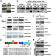
AGO1 is a peripheral membrane protein. (A) Western analysis of AGO1, HMG1, and PEPC proteins in 100,000 × g supernatant (sup) and pellet (pel) fractions of cleared Col-0 inflorescence extracts prepared by hypertonic lysis. Five-percent of the supernatant fraction is loaded, 20% of pellet fraction is loaded, precluding any clear estimates of relative abundance in soluble versus insoluble fractions. HMG1 is used here solely as a positive control for a transmembrane protein. (B) Western analysis of AGO1 and HMG1 proteins in pellet fractions prepared as in A. Pellets were resuspended in either microsome buffer, or microsome buffer supplemented with 1 M KCl, 0.1 M Na2CO3, or 0.5% Triton X-100, and separated into supernatant (sup) and pellet (pel) fractions again at 100,000 × g. At longer exposures, an insoluble AGO1 fraction upon Triton X-100 treatment is visible. (C) Microsome fractionation of inflorescence lysates from Col-0, ago1-25, and ago1-38. (Top) Western analysis of AGO1 in total extracts. (Middle) Western analysis of AGO1 in equally loaded microsome fractions, developed by enhanced chemiluminescence. (Bottom) Same analysis as in the Middle, developed by less sensitive alkaline phosphatase staining that allows a clearer visualization of the difference in microsomal AGO1 abundance between Col-0 (WT) and ago1-38. (D) (Upper) Schematic depicting the position of several AGO1 hypomorphic mutations used in this study (black triangle) and the ago1-38 allele (red triangle) causing the G186R missense mutation. (Lower) The glycine residue mutated in Arabidopsis ago1-38 is highly conserved among various metazoan and fungal AGO proteins (highlighted in yellow). At, Arabidopsis thaliana; Ce, Caenorhabditis elegans; Hs, Homo sapiens; Sp, Schizosaccharomyces pombe. (E) Microsome fractionation of inflorescence lysates from Col-0 and hmg1-3. (Upper) Western analysis of AGO1 in total extracts. (Lower) Western analysis of AGO1 and HMG1 in microsome (pel) fraction. (F) Same analysis as in E performed with GFP171.1 and mad3 inflorescence lysates.

References

    1. Voinnet O. Origin, biogenesis and activity of plant microRNAs. Cell. 2009;136:669–687. - PubMed
    1. Bartel DP. MicroRNAs: Target recognition and regulatory functions. Cell. 2009;136:215–233. - PMC - PubMed
    1. Cikaluk DE, et al. GERp95, a membrane-associated protein that belongs to a family of proteins involved in stem cell differentiation. Mol Biol Cell. 1999;10:3357–3372. - PMC - PubMed
    1. Gibbings DJ, Ciaudo C, Erhardt M, Voinnet O. Multivesicular bodies associate with components of miRNA effector complexes and modulate miRNA activity. Nat Cell Biol. 2009;11:1143–1149. - PubMed
    1. Lee YS, et al. Silencing by small RNAs is linked to endosomal trafficking. Nat Cell Biol. 2009;11:1150–1156. - PMC - PubMed

Publication types