. 2012 Mar 13;109(11):E630-9.
doi: 10.1073/pnas.1111303109. Epub 2012 Jan 13.

Alternative reading frame selection mediated by a tRNA-like domain of an internal ribosome entry site

Affiliations

Alternative reading frame selection mediated by a tRNA-like domain of an internal ribosome entry site

Qian Ren et al. Proc Natl Acad Sci U S A. .

Abstract

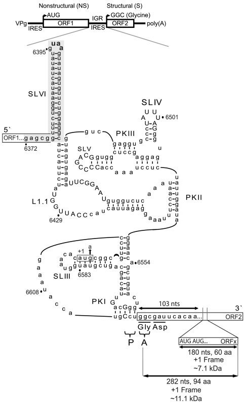
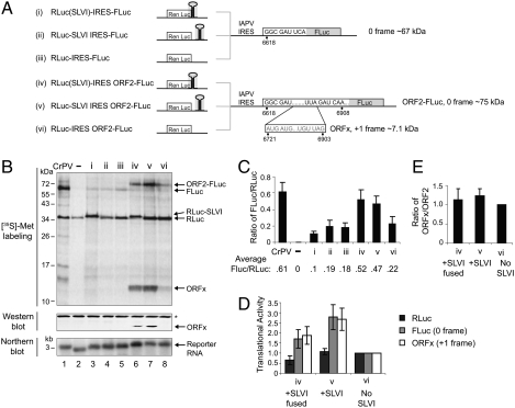
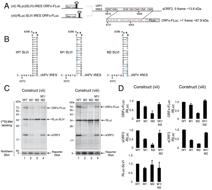
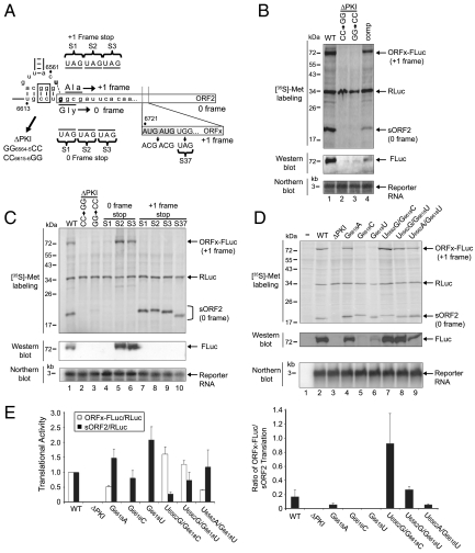
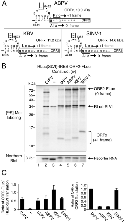
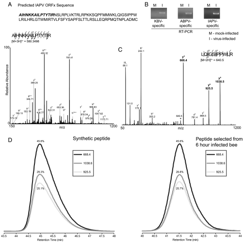
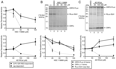
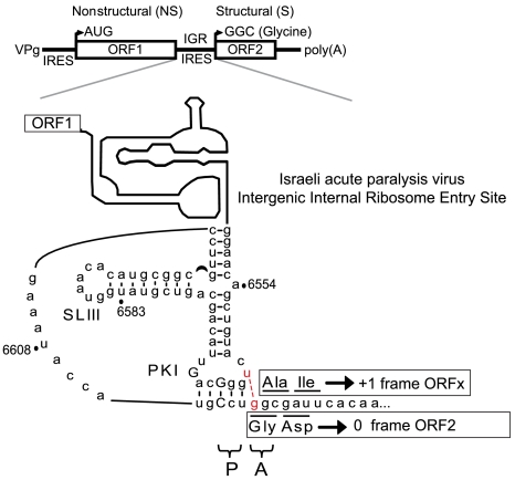
The dicistrovirus intergenic region internal ribosome entry site (IRES) utilizes a unique mechanism, involving P-site tRNA mimicry, to directly assemble 80S ribosomes and initiate translation at a specific non-AUG codon in the ribosomal A site. A subgroup of dicistrovirus genomes contains an additional stem-loop 5'-adjacent to the IRES and a short open reading frame (ORFx) that overlaps the viral structural polyprotein ORF (ORF2) in the +1 reading frame. Using mass spectrometry and extensive mutagenesis, we show that, besides directing ORF2 translation, the Israeli acute paralysis dicistrovirus IRES also directs ORFx translation. The latter is mediated by a UG base pair adjacent to the P-site tRNA-mimicking domain. An ORFx peptide was detected in virus-infected honey bees by multiple reaction monitoring mass spectrometry. Finally, the 5' stem-loop increases IRES activity and may couple translation of the two major ORFs of the virus. This study reveals a novel viral strategy in which a tRNA-like IRES directs precise, initiator Met-tRNA-independent translation of two overlapping ORFs.


Conflict of interest statement

The authors declare no conflict of interest.

Figures

Fig. 1.
Fig. 1.
Secondary structure of IAPV IGR IRES. (Top) Schematic of IAPV genome. Distinct IRESs direct translation of nonstructural (ORF1) and structural (ORF2) polyproteins. Schematic of IAPV IGR IRES showing pseudoknots PKI, PKII, and PKIII, stem loops SLIII, SLIV, SLV, and SLVI (shaded gray), and loop L1.1. The UAA stop codon of ORF1 is shown in bold within the loop of SLVI. The overlapping +1 frame ORFx, within ORF2, is shown. Tandem ORFx-frame AUG codons, 103 nucleotides downstream from the IGR IRES, are indicated and, if utilized as the initiation site for ORFx translation, would lead to a 7.1-kDa protein. If, on the other hand, ORFx translation initiates at or near the IGR IRES, then an 11.1-kDa protein is expected. AUG6592-4 (boxed) is an alternative potential initiation site for +1 frame ORFx. G6594A tests whether the AUG6592-4 codon is used for ORFx translation. Conserved nucleotides among type II IGR IRESs are in capital letters. The CCU triplet mediates PKI base pairing and occupies the P site, whereas the first codon of the 0 frame ORF2 is the 3′-adjacent GGC codon in the A site.
Fig. 2.
Fig. 2.
Characterization of novel features within the IAPV IGR IRES. (A) Schematic of dicistronic reporter constructs containing the IAPV IGR IRES inserted in the intergenic region between two reporter genes: Renilla luciferase (RLuc), which monitors scanning-mediated translation, and firefly luciferase (FLuc), which monitors IRES-mediated translation. Constructs (i)–(iii) contain the IAPV IGR IRES alone. Constructs (iv)–(vi) also contain a part of the IAPV ORF2 (6618–6908) fused in the 0 frame with FLuc (ORF2-FLuc). This region also contains the predicted ORFx in the +1 frame. Dicistronic reporter constructs were engineered to contain no SLVI (iii, vi), SLVI positioned in the IGR (ii, v), or SLVI positioned such that the UAA stop codon within SLVI serves as the termination codon for RLuc (i, iv). (B) The presence of SLVI and part of ORF2 stimulates IAPV IGR IRES translation. Dicistronic reporter constructs were incubated in Sf21 extracts at 30 °C for 120 min in the presence of [35S]-methionine. CrPV and (−) denote dicistronic reporters containing the CrPV IGR IRES and an empty no-IRES control, respectively. In parallel, reactions were subjected to Western blotting, or to Northern blotting using a probe specific to the reporter RNA. (C) Quantitation of radiolabeled protein products calculated by taking the ratio FLuc/RLuc. Averages from at least three independent experiments ± SD are shown. (D) SLVI increases IGR IRES activity. Graph of translational activities determined via quantitation of radiolabeled proteins produced from constructs (iv), (v), and (vi). Values were normalized relative to (vi). (E) Ratio of ORFx/ORF2 protein products produced from the indicated constructs. All calculations are from at least three independent experiments ± SD.
Fig. 3.
Fig. 3.
The integrity of SLVI affects IAPV IGR IRES translation. (A) Schematic of dicistronic constructs containing the IAPV IGR IRES and ORFx fused in frame with FLuc. Red nucleotides denote mutations inserted to fuse the +1 frame ORFx in frame with FLuc. These mutations also create a short 0 frame ORF2 protein (sORF2). (B) Schematic of WT and mutant (M1 and M2) SLVI. Mutations that disrupt the helical base pairing within SLVI (M1 and M2) are indicated in blue. Compensatory mutations (M1/M2 combined) restore base pairing within SLVI. Bold nucleotides denote the UAA stop codon of ORF1. (C) Dicistronic constructs containing IGR SLVI (viii) or SLVI fused with RLuc (vii) were incubated in Sf21 translation extracts. Northern blotting using a probe specific to the reporter RNA is shown. (D) Quantitation of translation products normalized relative to the dicistronic construct containing the wild-type SLVI. Averages ± SD are from at least three independent experiments.
Fig. 4.
Fig. 4.
U6562/G6618 base pairing directs +1 frame ORFx translation. (A) Schematic of the IAPV PKI domain. Black dashes and bold denote the predicted U6562/G6618 base pairing within PKI which directs +1 frame ORFx translation. The first amino acid of ORF2 is encoded by a glycine GGC codon in the 0 frame. The predicted first amino acid of ORFx—if ORFx is initiated at the IGR IRES—is encoded by an alanine GCG codon in the +1 frame. Tandem AUG codons within ORFx are shown. Mutations that replace codons with a stop codon in the 0 or +1 frame are denoted by S1-3 and S37. Mutations that disrupt PKI base pairing are shown (ΔPKI). Mutations were inserted in reporter constructs (viii). (BD) Reporter constructs were incubated in Sf21 extracts. In parallel, reactions were subjected to Western blotting or Northern blotting. “comp” denotes compensatory mutations to restore PKI base pairing. (E) Quantitation of radiolabeled protein products normalized relative to the wild-type IAPV IGR IRES dicistronic reporter is shown (Left). The ratio of ORFx-FLuc (+1 frame)/sORF2 (0 frame) translation, taking into account the number of methionines within each translated protein, is shown (right). Averages ± SD are from at least three independent experiments.
Fig. 5.
Fig. 5.
A subset of dicistrovirus IGR IRESs direct ORFx translation. (A) Schematic of the PKI domains of the subset of dicistroviruses that contain the +1 frame ORFx. Black dashes denote the predicted additional base pairing within PKI which may direct +1 frame ORFx translation. (B) Dicistronic reporter constructs (iv) containing the KBV, ABPV, or SINV-1 IGR IRESs were incubated in Sf21 extracts. A representative SDS-PAGE of radiolabeled protein products detected by autoradiography is shown. In parallel, reactions were subjected to Northern blotting. (C) Quantitation of radiolabeled protein products normalized relative to the wild-type IAPV IGR IRES dicistronic reporter is shown at top. +1 frame ORFx or 0 frame sORF2-FLuc translated proteins were normalized to RLuc. The ratio of ORFx/sORF2-FLuc translation, taking into account the number of methionines within each translated protein, is shown. Averages ± SD are from at least three independent experiments.
Fig. 6.
Fig. 6.
Detection of ORFx in vitro and in virus-infected honey bee pupae. (A) Identification of the start site of ORFx. Fragment spectra from the 580.3498 Th, triply charged precursor ion of AIHNKKAILPTYTIR from ArgC-digested ORFx indicating that ORFx translation starts at this Ala residue. Individual fragment ions are annotated in the spectrum and in the sequence representation. (B) Detection of KBV, ABPV, and IAPV from virus-infected honey bee pupae by RT-PCR using virus-specific primers. RNA was extracted from honey bee pupae 96 h after injection with PBS (mock-infected) or virus particles. (C) Fragment spectra from a shared peptide of KBV and IAPV ORFx with the three y ions selected for transitions labeled in bold. (D) MRM traces for the pure synthetic peptide and the same three transitions detected in virally infected bees at 6 h after injection. The relative percentages of area under the curve for each transition are labeled.
Fig. 7.
Fig. 7.
The 0 and +1 frame translation when eIF2 or eIF4F activity is compromised in vitro. Capped in vitro transcribed dicistronic reporter RNAs containing the (A) CrPV IGR IRES or (B and C) IAPV IGR IRES (construct iv) were incubated in Sf21 extracts alone, with DMSO or with NSC119889 or 4E1RCat, which inhibit eIF2 activity or eIF4E-eIF4G interactions, respectively. Extracts were preincubated for 5 min with the compound prior to adding the reporter RNA. Shown are the relative quantitation of the cap-dependent (RLuc), 0 frame (ORF2-FLuc), and +1 frame (ORFx) translation radiolabeled protein products. All calculations are from at least three independent experiments ± SD.
Fig. P1.
Fig. P1.
Schematic of the honey bee Israeli acute paralysis virus genome (Top) and secondary structure of the IAPV intergenic internal ribosome entry site (intergenic region IRES). Shown is the tRNA-mimic pseudoknot I (PKI) that occupies the ribosomal P site to direct translation initiation of the viral structural polyprotein ORF2 at a glycine GGC codon in the ribosomal A site. An extra U:G base pair (red nucleotides), adjacent to pseudoknot I, shifts the reading frame by +1 nucleotide to direct translation initiation of ORFx at an alanine GCG codon in the ribosomal A site. This study reveals a unique viral strategy in which the tRNA-like viral intergenic region IRES directs translation of two overlapping ORFs.

References

    1. Bonning BC, Miller WA. Dicistroviruses. Annu Rev Entomol. 2010;55:129–150. - PubMed
    1. Wilson JE, Powell MJ, Hoover SE, Sarnow P. Naturally occurring dicistronic cricket paralysis virus RNA is regulated by two internal ribosome entry sites. Mol Cell Biol. 2000;20:4990–4999. - PMC - PubMed
    1. Cox-Foster DL, et al. A metagenomic survey of microbes in honey bee colony collapse disorder. Science. 2007;318:283–287. - PubMed
    1. Jan E, Kinzy TG, Sarnow P. Divergent tRNA-like element supports initiation, elongation, and termination of protein biosynthesis. Proc Natl Acad Sci USA. 2003;100:15410–15415. - PMC - PubMed
    1. Jan E, Sarnow P. Factorless ribosome assembly on the internal ribosome entry site of cricket paralysis virus. J Mol Biol. 2002;324:889–902. - PubMed

Publication types

LinkOut - more resources