Review
. 2012 Feb;54 Suppl 1(Suppl 1):S8-S15.
doi: 10.1093/cid/cir864.

Taxonomy of fungi causing mucormycosis and entomophthoramycosis (zygomycosis) and nomenclature of the disease: molecular mycologic perspectives

Affiliations
Review

Taxonomy of fungi causing mucormycosis and entomophthoramycosis (zygomycosis) and nomenclature of the disease: molecular mycologic perspectives

Kyung J Kwon-Chung. Clin Infect Dis. 2012 Feb.

Abstract

Molecular phylogenetic analysis confirmed the phylum Zygomycota to be polyphyletic, and the taxa conventionally classified in Zygomycota are now distributed among the new phylum Glomeromycota and 4 subphyla incertae sedis (uncertain placement). Because the nomenclature of the disease zygomycosis was based on the phylum Zygomycota (Zygomycetes) in which the etiologic agents had been classified, the new classification profoundly affects the name of the disease. Zygomycosis was originally described as a convenient and inclusive name for 2 clinicopathologically different diseases, mucormycosis caused by members of Mucorales and entomophthoramycosis caused by species in the order Entomophthorales of Zygomycota. Without revision of original definition, the name "zygomycosis," however, has more often been used as a synonym only for mucormycosis. This article reviews the progress and changes in taxonomy and nomenclature of Zygomycota and the disease zygomycosis. The article also reiterates the reasons why the classic names "mucormycosis" and "entomophthoramycosis" are more appropriate than "zygomycosis."


Figures

Figure 1.
Figure 1.
Old (A) and a proposed new (B) classification schemes of the kingdom Fungi.
Figure 2.
Figure 2.
Phylogeny of fungi determined on the basis of small subunit ribosomal RNA sequences. Thick lines delineate clades supported by bootstrap values >90%. “Zygomycota” do not form monophyletic clades [9]. The metric bar indicates number of substitutions per site.
Figure 3.
Figure 3.
Phylogenetic tree provided by T. Y. James at the University of Michigan, Ann Arbor, which was based on unpublished results from the AFTOL (Assembling the Fungal Tree of Life) project [10]. Results are similar to those reported by James et al [5], but additional basal taxa are included.
Figure 4.
Figure 4.
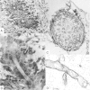
Morphology of conidia and zygospores (scale bar, 20 μm). A, Reproduction of a conidium in Basidiobolus ranarum. A conidium discharged onto a Petri dish cover germinated and produced a conidiophore bearing a single conidium. B, Primary conidia of Conidiobolus incongruus germinated to produce long hyphae bearing subglobose conidia [38]. C, Secondary conidium formation by replication in Conidiobolus coronatus. D, Zygospore of Basidiobolus ranarum, with a characteristic beak, is produced by the fusion of 2 adjacent hyphal cells. E, Sporangial structure of Rhizopus species showing the sporangiophore (S), apophysis (A), columella (C), and sporangiospores (SP). F, Sporangium of Lichtheimia (Absidia) corymbifera. G, Electron microscopy of Zygospore (ZS) of Rhizopus species produced between 2 suspensors (S) originating from hyphae of 2 sexually compatible strains (courtesy of Dr S. L. Flegler). Images in A and C–F are from Kwon-Chung and Bennett [33].
Figure 5.
Figure 5.
A, Basidiobolus ranarum in hematoxylin-eosin–stained tissue section showing a cross-section of hyphae with sleeves of eosinophilic material. B, Hyphae in longitudinal section in the same tissue shown in A. C, Angioinvasion in mucormycosis. A pulmonary blood vessel occluded by hyphae of Cunninghamella bertholletiae stained with hematoxylin-eosin. D, Hyphae of Rhizopus species in tissue stained by hematoxylin-eosin. Note the lack of eosinophilic material around the hyphae. Images are reproduced from Kwon-Chung and Bennett [33] (scale bar, 10 μm).

References

    1. Whittaker RH. New concepts of kingdoms of organisms. Evolutionary relations are better represented by new classifications than by the traditional two kingdoms. Science. 1969;163:150–60. - PubMed
    1. Emmons CW, Binford CH, Utz JP, Kwon-Chung KJ. Medical mycology. 3rd ed. Philadelphia: Lea & Febiger; 1977. pp. 254–84.
    1. Ainsworth GC. Introduction and keys to higher taxa. In: Ainsworth GC, Sparrow FK, Sussman AS, editors. The fungi. IVA. A taxonomic review with keys. New York: Academic Press; 1973. pp. 1–7.
    1. Kirk PM, Cannon PF, David JC, Stalpers JA. Dictionary of the fungi. 9th ed. Wallingford, United Kingdom: CAB International; 2001.
    1. James TY, Kauff F, Schoch CL, et al. Reconstructing the early evolution of fungi using a six-gene phylogeny. Nature. 2006;443:818–22. - PubMed

MeSH terms