Comparative Study
. 2012 Feb 23;53(2):959-66.
doi: 10.1167/iovs.11-8377. Print 2012 Feb.

Myeloid suppressor cells induced by retinal pigment epithelial cells inhibit autoreactive T-cell responses that lead to experimental autoimmune uveitis

Affiliations
Comparative Study

Myeloid suppressor cells induced by retinal pigment epithelial cells inhibit autoreactive T-cell responses that lead to experimental autoimmune uveitis

Zhidan Tu et al. Invest Ophthalmol Vis Sci. .

Abstract

Purpose: To test whether retinal pigment epithelial (RPE) cells are able to induce myeloid-derived suppressor cell (MDSC) differentiation from bone marrow (BM) progenitors.

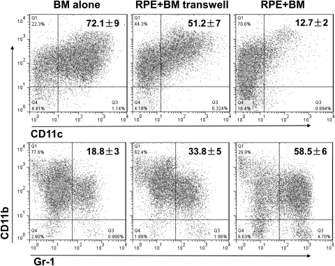
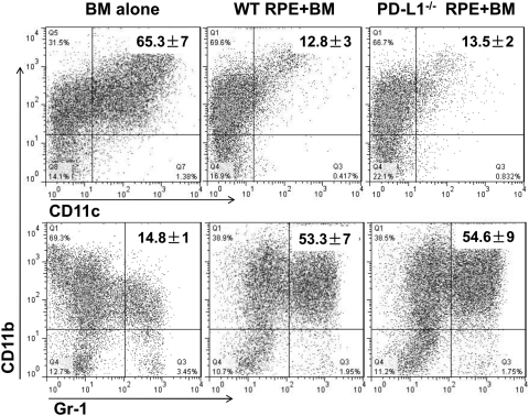
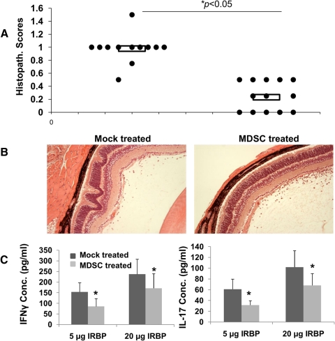
Methods: BM cells were cocultured with or without RPE cells in the presence of GM-CSF and IL-4. Numbers of resultant MDSCs were assessed by flow cytometry after 6 days of incubation. The ability of the RPE cell-induced MDSCs to inhibit T cells was evaluated by a CFSE-based T-cell proliferation assay. To explore the mechanism by which RPE cells induce MDSC differentiation, PD-L1-deficient RPE cells and blocking antibodies against TGF-β, CTLA-2α, and IL-6 were used. RPE cell-induced MDSCs were adoptively transferred into mice immunized with interphotoreceptor retinoid-binding protein in complete Freund's adjuvant to test their efficacy in suppressing autoreactive T-cell responses in experimental autoimmune uveitis (EAU).

Results: RPE cells induced the differentiation of MDSCs. These RPE cell-induced MDSCs significantly inhibited T-cell proliferation in a dose-dependent manner. PD-L1-deficient RPE cells induced MDSC differentiation as efficiently as wild-type RPE cells, and neutralizing TGF-β or CTLA-2α did not alter the numbers of induced MDSCs. However, blocking IL-6 reduced the efficacy of RPE cell-induced MDSC differentiation. Finally, adoptive transfer of RPE cell-induced MDSCs suppressed IRBP-specific T-cell responses that led to EAU.

Conclusions: RPE cells induce the differentiation of MDSCs from bone marrow progenitors. Both cell surface molecules and soluble factors are important in inducing MDSC differentiation. PD-L1, TGF-β, and CTLA-2α were not measurably involved in RPE cell-induced MDSC differentiation, whereas IL-6 was important in the process. The induction of MDSCs could be another mechanism by which RPE cells control immune reactions in the retina, and RPE cell-induced MDSCs should be further investigated as a potential approach to therapy for autoimmune posterior uveitis.


Figures

Figure 1.
Figure 1.
RPE cells inhibited DC propagation and induced MDSC differentiation. BM cells were cultured without and with RPE (at a ratio of 20:1) in the presence of GM-CSF and IL-4. After incubation, nonadherent cells were analyzed for markers of DCs (CD11b+CD11c+) (A) and MDSCs (CD11b+GR-1+) (B). Photographs of the resultant DCs (C) and MDSC (D) were taken after conventional Giemsa staining. Results are representative of more than individual experiments. Data are mean ± SD.
Figure 2.
Figure 2.
RPE cell–induced MDSCs inhibited T-cell responses. Spleen cells (5 × 10⁵) from naive C57BL/6 mice were labeled with CFSE and activated by anti–CD3 mAb, then cocultured with different numbers of the RPE cell–induced MDSCs. In 2 days, the inhibition of T-cell responses was assessed by evaluating proliferating T cell–formed clusters directly under a microscope (A, top) and by measuring CFSE dilution using flow cytometry, gating on the CD4+ T cells (A, bottom). The inhibition of T-cell responses (B) was calculated using the following formula: inhibition (%) = 1 − [(ba)/a], where a is the number of proliferating T cells without MDSCs and b is the number of proliferating T cells with MDSCs. IFN-γ levels in the supernatants were measured by standard ELISA (C).
Figure 3.
Figure 3.
Both RPE cell surface molecules and RPE cell–derived soluble factors were involved in suppressing DC propagation and inducing MDSC differentiation. BM cells were cultured without and with RPE (at a ratio of 20:1) in direct contact or in culture inserts together with GM-CSF and IL-4. After incubation, nonadherent cells were analyzed for markers of DCs (CD11b+CD11c+) and MDSCs (CD11b+GR-1+). Results are representative of two individual experiments. Data are mean ± SD.
Figure 4.
Figure 4.
The cell surface molecule PD-L1 was not involved in RPE cell–induced MDSC differentiation. BM cells were cultured without and with WT or PD-L1−/− RPE (at a ratio of 20:1), together with GM-CSF and IL-4. After incubation, nonadherent cells were analyzed for markers of DCs (CD11b+CD11c+) and MDSCs (CD11b+GR-1+). Results are representative of two individual experiments. Data are mean ± SD.
Figure 5.
Figure 5.
The soluble factor IL-6 is important for RPE cell–induced MDSC differentiation. BM cells were cultured without and with RPE (at a ratio of 20:1), together with GM-CSF and IL-4 for 3 days. Then supernatants were collected, and concentrations of IL-6 were measured by ELISA (A). The same experiments were repeated except that in half the wells, 5 μg/mL anti–IL-6 IgG was added. In the other half, the same amount of isotype control was added. After incubation, nonadherent cells were analyzed for markers of MDSC (CD11b+GR-1+). Results are representative of two individual experiments. Data are mean ± SD. *P < 0.05.
Figure 6.
Figure 6.
RPE cell–induced MDSCs suppress in vivo autoreactive T-cell responses that lead to retinal injury in EAU. RPE cell–induced MDSCs (2 × 10⁶) in 0.5 mL PBS were adoptively transferred into each of the six female C57BL/6 mice after EAU induction by tail vein intravenous injection. The same volume of PBS was administered into each of the six control mice. In 21 days, EAU disease severity was evaluated by histopathologic scoring of the ocular sections in a masked fashion. Each dot represents one eye (A). Shown are representative images of the retina from the mock-treated and MDSC-treated mice (B). IRBP-specific Th1 and Th17 cell responses in the experimental mice were assessed by IFN-γ and IL-17 ELISA using culture supernatants collected from splenocytes restimulated with different concentrations of the IRBP1–20 peptide (C).

References

    1. Young MR, Newby M, Wepsic HT. Hematopoiesis and suppressor bone marrow cells in mice bearing large metastatic Lewis lung carcinoma tumors. Cancer Res. 1987;47:100–105 - PubMed
    1. Buessow SC, Paul RD, Lopez DM. Influence of mammary tumor progression on phenotype and function of spleen and in situ lymphocytes in mice. J Natl Cancer Inst. 1984;73:249–255 - PubMed
    1. Seung LP, Rowley DA, Dubey P, Schreiber H. Synergy between T-cell immunity and inhibition of paracrine stimulation causes tumor rejection. Proc Natl Acad Sci U S A. 1995;92:6254–6258 - PMC - PubMed
    1. Kusmartsev S, Nefedova Y, Yoder D, Gabrilovich DI. Antigen-specific inhibition of CD8+ T cell response by immature myeloid cells in cancer is mediated by reactive oxygen species. J Immunol. 2004;172:989–999 - PubMed
    1. Cripps JG, Gorham JD. MDSC in autoimmunity. Int Immunopharmacol. 2011;11:789–793 - PMC - PubMed

Publication types