Comparative Study
. 2012 Jan 31;53(1):501-7.
doi: 10.1167/iovs.11-8784.

Generation of an inbred miniature pig model of retinitis pigmentosa

Affiliations
Comparative Study

Generation of an inbred miniature pig model of retinitis pigmentosa

Jason W Ross et al. Invest Ophthalmol Vis Sci. .

Abstract

Purpose: The Pro23His (P23H) rhodopsin (RHO) mutation underlies the most common form of human autosomal dominant retinitis pigmentosa (adRP). The objective of this investigation was to establish a transgenic miniature swine model of RP using the human P23H RHO gene.

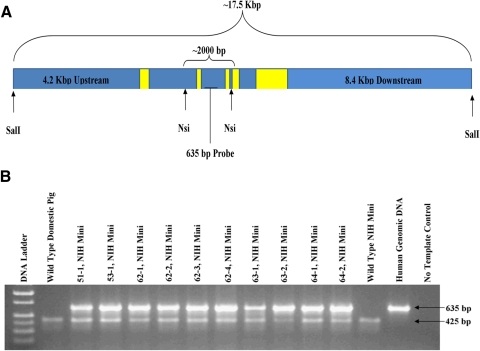
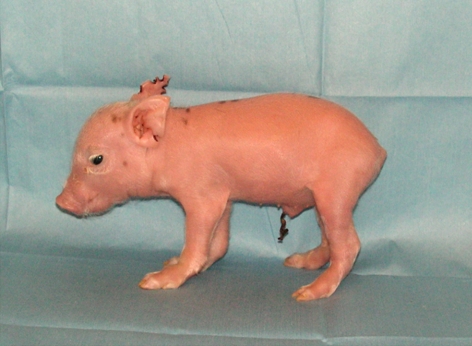
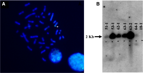
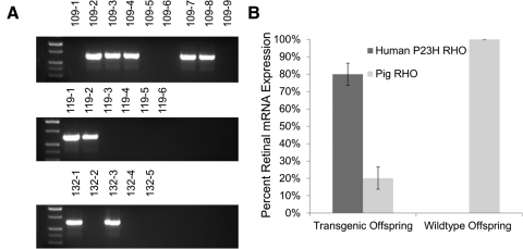
Methods: Somatic cell nuclear transfer (SCNT) was used to create transgenic miniature pigs that expressed the human P23H RHO mutation. From these experiments, six transgenic founders were identified whose retinal function was studied with full-field electroretinography (ffERG) from 3 months through 2 years. Progeny from one founder were generated and genotyped to determine transgene inheritance pattern. Retinal mRNA was isolated, and the ratio of P23H to wild-type pig RHO was measured.

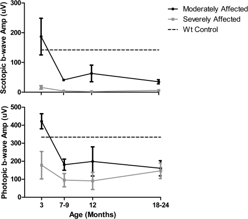
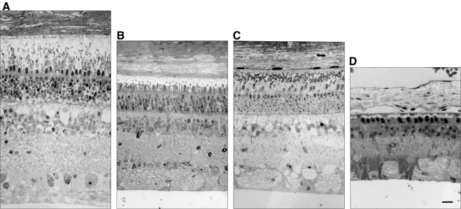
Results: A single transgene integration site was observed for five of the six founders. All founders had abnormal scotopic and photopic ffERGs after 3 months. The severity of the ffERG phenotype was grouped into moderately and severely affected groups. Offspring of one founder inherited the transgene as an autosomal dominant mutation. mRNA analyses demonstrated that approximately 80% of total RHO was mutant P23H.

Conclusions: Expression of the human RHO P23H transgene in the retina creates a miniature swine model with an inheritance pattern and retinal function that mimics adRP. This large-animal model can serve as a novel tool for the study of the pathogenesis and therapeutic intervention in the most common form of adRP.


Figures

Figure 1.
Figure 1.
Generation of P23H miniature pig RP model. (A) The 17.5-kb construct provided by Thaddeus Dryja. The construct was linearized with SalI before electroporation into the cells. Yellow boxes: approximate location and size of the five RHO exons. NsiI was used for Southern blot analysis detected by a 635-bp radiolabeled probe. (B) A total of 10 piglets were born alive, all PCR positive for the genomic human RHO construct (635-bp amplicon), while endogenous porcine gDNA (425-bp amplicon) was amplified in all pig samples.
Figure 2.
Figure 2.
The first of six founder piglets, 51-1, born May 25, 2008.
Figure 3.
Figure 3.
(A) A representative FISH image (founder 53-1) indicating the chromosomal integration site of the human RHO transgene. All founder pigs demonstrated a single integration site, except 51-1 for which a signal was not detected. (B) Southern blot of six living founders. A total of 10 μg digested with NsiI was loaded per lane. Pig 18-1 was the original source of the fetal fibroblasts used to create the transgenic founders. Signal for all transgenic pigs was detected, while 51-1 and 64-1 demonstrated the least signal. Pigs 53-1 and 63-2 most likely had a greater number of human RHO copies integrated into the genome.
Figure 4.
Figure 4.
Time course of decrease in retinal function differs in P23H transgenic minipig founders. (A) Scotopic (rod) and (B) photopic (cone) full-field ERGs were recorded during the first 2 years of life under standard ISCEV protocols. The scotopic ERGs were evoked with a flash intensity of 0.01 cd · s · m−2, after 20 minutes of dark-adaptation. The photopic ERGs were measured using flashes of 3 cd · s · m−2 presented on a background of 30 cd · m−2. The founders fell into two groups, based on both scotopic and photopic ffERGs, compared with WT littermates at 3 months of age (n = 3; dashed lines). Average b-wave amplitudes are plotted for the two transgenic founders who were moderately affected (63-1 and 51-1) and the four who were severely affected (53-1, 62-1, 63-2, and 64-1). Moderately affected founders had a later onset of rod and cone dysfunction and retained some degree of both rod and cone response through 12 months of age.
Figure 5.
Figure 5.
Retinal histology of three of six P23H founders at death. (A) Retinal morphology of a normal adult domestic sow. (B) 51-1 (18 months) and (C) 63-1 (22 months). The outer segments of the photoreceptors were shortened in the moderately affected ffERG group in the two founders, and the ONL thickness was reduced compared with that in the normal adult. (D) 63-2 (12 months). Only a single layer of cone nuclei remained in the ONL and both the ONL and INL were greatly reduced in thickness in the founder with a severely affected ffERG. Thus, there is a correlation between retinal structure and function in the P23H model. Scale bar, 50 μm.
Figure 6.
Figure 6.
Characterization of F1 progeny produced by 53-1. (A) Three litters were produced from founder 53-1 after mating to a domestic pig (litter 109) and two miniature pigs (litters 118 and 132). PCR screening of the 20 offspring detected 9 transgenic piglets, suggesting normal Mendelian inheritance. Controls included genomic DNA from a wild-type miniature pig (WT-1), a wild-type domestic pig (WT-2), human genomic DNA, and a no-template control (NTC). Transgenic positive amplicon is 635 bp. (B) Contribution of both human P23H and pig RHO to the total RHO mRNA expression in the neonatal retina of transgenic (109-2, -3, -4, -7, and -8) and wild-type (109-1, -5, -6, and -9) offspring. Retinal RNA extraction, followed by PCR amplification, cloning, and sequencing, identified the mRNA expression ratio of endogenous porcine RHO mRNA versus the transgenic human P23H RHO mRNA.

Comment in

Similar articles

Cited by

References

    1. Inglehearn CF. Molecular genetics of human retinal dystrophies. Eye (Lond). 1998;12:571–579 - PubMed
    1. Simonelli F, Maguire AM, Testa F, et al. Gene therapy for Leber's congenital amaurosis is safe and effective through 1.5 years after vector administration. Mol Ther. 18:643–650 - PMC - PubMed
    1. MacLaren RE, Pearson RA, MacNeil A, et al. Retinal repair by transplantation of photoreceptor precursors. Nature. 2006;444:203–207 - PubMed
    1. Hendrickson A, Hicks D. Distribution and density of medium- and short-wavelength selective cones in the domestic pig retina. Exp Eye Res. 2002;74:435–444 - PubMed
    1. Inglehearn CF, Tarttelin EE, Plant C, et al. A linkage survey of 20 dominant retinitis pigmentosa families: frequencies of the nine known loci and evidence for further heterogeneity. J Med Genet. 1998;35:1–5 - PMC - PubMed

Publication types

MeSH terms