. 2012 Mar;194(6):1523-32.
doi: 10.1128/JB.06104-11. Epub 2012 Jan 13.

Characterization of Escherichia coli dinJ-yafQ toxin-antitoxin system using insights from mutagenesis data

Affiliations

Characterization of Escherichia coli dinJ-yafQ toxin-antitoxin system using insights from mutagenesis data

Julija Armalyte et al. J Bacteriol. 2012 Mar.

Abstract

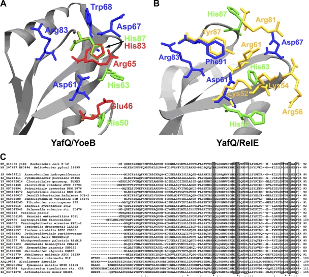
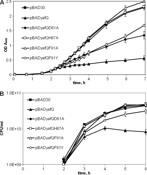
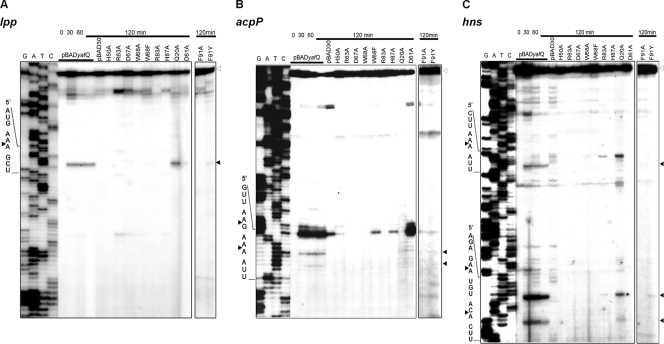
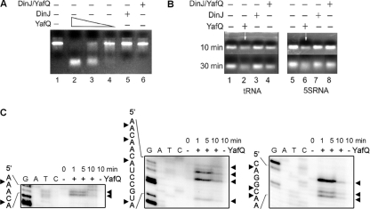
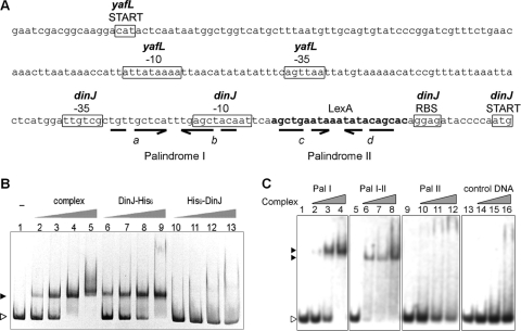
Escherichia coli dinJ-yafQ operon codes for a functional toxin-antitoxin (TA) system. YafQ toxin is an RNase which, upon overproduction, specifically inhibits the translation process by cleaving cellular mRNA at specific sequences. DinJ is an antitoxin and counteracts YafQ-mediated toxicity by forming a strong protein complex. In the present study we used site-directed mutagenesis of YafQ to determine the amino acids important for its catalytic activity. His50Ala, His63Ala, Asp67Ala, Trp68Ala, Trp68Phe, Arg83Ala, His87Ala, and Phe91Ala substitutions of the predicted active-site residues of YafQ abolished mRNA cleavage in vivo, whereas Asp61Ala and Phe91Tyr mutations inhibited YafQ RNase activity only moderately. We show that YafQ, upon overexpression, cleaved mRNAs preferably 5' to A between the second and third nucleotides in the codon in vivo. YafQ also showed RNase activity against mRNA, tRNA, and 5S rRNA molecules in vitro, albeit with no strong specificity. The endoribonuclease activity of YafQ was inhibited in the complex with DinJ antitoxin in vitro. DinJ-YafQ protein complex and DinJ antitoxin alone selectively bind to one of the two palindromic sequences present in the intergenic region upstream of the dinJ-yafQ operon, suggesting the autoregulation mode of this TA system.


Figures

Fig 1
Fig 1
YafQ active-site prediction. (A) Active site of YafQ structural model, created using YoeB toxin structure (PDB code 2a6s). (B) Active site of YafQ structural model, created using RelE from E. coli (PDB code 3kha). The amino acids important for YoeB catalysis are drawn in red and for RelE are drawn in yellow. The absolutely conservative histidines of YafQ are drawn in green; other conservative or partially conservative amino acids of YafQ are in blue. The structure models of YafQ were created by the Swiss-Model automated protein homology modeling server (1). (C) Sequence alignment of YafQ homologues from different bacteria. Absolutely conserved histidines are indicated by gray boxes; other possibly important active-site amino acids are indicated by boxes. Identical amino acid residues are indicated by asterisks below, and similar amino acid residues are indicated with colons or dots below. Sequences were aligned using CLUSTAL W, v1.82.
Fig 2
Fig 2
Growth of E. coli BW25113 after induction of YafQ mutant proteins. Cells with pBADyafQ, pBAD30 and pBADyafQ plasmids with Gln20Ala, Asp61Ala, His87Ala, Phe91Ala, and Phe91Tyr mutations in yafQ gene were grown in liquid LB medium to an A600 of 0.1 as described in Materials and Methods. Then synthesis of the proteins was induced by adding 0.2% of l-arabinose (2-h time point). The growth was monitored at the selected time points as the OD600 (A) or the CFU/ml (B). The data are means of at least three independent experiments. Bars indicate the standard error.
Fig 3
Fig 3
Primer extension analysis of cellular transcripts after induction of YafQ mutant proteins in vivo. BW25113 strain containing plasmids pBADyafQ, pBAD30, or pBADyafQ with Gln20Ala, His50Ala, Asp61Ala, His63Ala, Asp67Ala, Trp68Ala, Trp68Phe, Arg83Ala, His87Ala, Phe91Ala, and Phe91Tyr substitutions were grown to mid-exponential phase, and the synthesis of YafQ was induced by adding 0.2% of arabinose as described in Materials and Methods. At selected time points, the total RNA was extracted and used for primer extension reactions with lpp-, acpP-, and hns-specific 5′-32P-labeled primers. The sequence ladder was obtained by dideoxy DNA sequencing reactions using the corresponding primers for primer extension. The cleavage sites of the YafQ toxin are indicated by full arrowheads, and the full-length RNAs are indicated by empty arrowheads.
Fig 4
Fig 4
Analysis of RNA cleavage by YafQ toxin in vitro. (A) In vitro-synthesized substrate (asr) mRNA (7.5 μg) was incubated for 30 min in 37°C in 50 mM Tris-HCl (pH 7.0) with, respectively, 0.5, 0.25, and 0.1 μM YafQ(His)6 (lanes 2 to 4), 0.5 μM DinJ(His)6 (lane 5), and 0.5 μM DinJ-YafQ(His)6 protein complex (lane 6). (B) 5S rRNA or tRNA (0.16 μg) (lanes 1 and 5) was denatured at 70°C for 10 min and then incubated for 30 min in 37°C in 50 mM Tris-HCl (pH 7.0) with 1 μM YafQ(His)6 (lanes 2 and 6), 1 μM DinJ(His)6 (lanes 3 and 7), 1 μM DinJ-YafQ(His)6 complex (lanes 4 and 8). The sample volume was 10 μl, and samples were analyzed in a 2% agarose gel, run for 10 or 30 min as indicated on the left (in panel B). (C) In vitro-synthesized substrate (hns) mRNA (0.2 μg) was incubated for 1, 5, or 10 min in 37°C in 50 mM Tris-HCl (pH 7.0) with 2 μM YafQ(His)6 (“+” lanes) or without YafQ(His)6 (“−” lanes); the sample volume was 5 μl. After incubation, the samples were heat inactivated and used for primer extension reactions with hns-specific 5′-32P-labeled primers. The sequence ladder was obtained by dideoxy DNA sequencing reactions with corresponding primers used for primer extension. The cleavage sites of the YafQ toxin are indicated by full arrowheads.
Fig 5
Fig 5
Analysis of dinJ-yafQ promoter region and its interaction with DinJ-YafQ(His)6 protein complex. (A) E. coli intergenic region containing dinJ-yafQ promoter and palindromic sequences. The important predicted (BPROM) promoter sequences of dinJ and yafL genes are boxed, the putative LexA binding site is indicated in boldface, broken arrows indicate palindromic sequences, and letters in italics indicate the palindrome sequence regions. (B) EMSA analysis of interaction of DinJ-YafQ(His)6 complex with 376-bp dinJ-yafQ promoter DNA fragment. A total of 8 pmol of PCR-amplified dinJ-yafQ promoter DNA fragments was incubated in 10 μl of binding buffer (10 mM Tris-HCl, 50 mM NaCl, 1 mM DTT, 5 mM MgCl2, 2.5% glycerol [pH 7.5]) with increasing amounts of protein at 22°C for 30 min. The EMSA was visualized in a 6% polyacrylamide gel stained with ethidium bromide. Lanes 1 to 5, promoter DNA incubated with 0, 0.5, 1, 2, and 4 pmol of DinJ-YafQ(His)6 protein complex; lanes 6 to 9, promoter DNA with 10, 20, 40, and 80 pmol of DinJ(His)6, lanes 10 to 13, promoter DNA with 10, 20, 40, and 80 pmol of (His)6DinJ. (C) EMSA analysis of DinJ-YafQ(His)6 protein complex interaction with the palindromic sequences of dinJ-yafQ promoter region. EMSA was performed to detect the interactions of purified DinJ-YafQ(His)6 protein complex with three 26-nucleotide regions of the dinJ-yafQ promoter, containing palindrome I (Pal I), 3′ part of palindrome I and the 5′ part of palindrome II (Pal I-II), and palindrome II (Pal II). Random DNA sequence was used as a control (control DNA). A total of 12.5 pmol of each DNA fragment (prepared as described in Materials and Methods) was incubated in 10 μl of binding buffer (10 mM Tris-HCl, 50 mM NaCl, 1 mM DTT, 5 mM MgCl2, 2.5% glycerol [pH 7.5]) with increasing amounts of protein (0, 8, 15, and 30 nmol) at 22°C for 30 min. EMSA results were visualized by ethidium bromide staining of an 8% polyacrylamide gel. The open arrowhead indicates free DNA; full arrowheads indicate DNA bound in complex with DinJ-YafQ(His)6.

References

    1. Arnold K, Bordoli L, Kopp J, Schwede T. 2006. The Swiss-Model Workspace: a web-based environment for protein structure homology modeling. Bioinformatics 22:195–201 - PubMed
    1. Bauerová-Hlinková V, Dvorský R, Perecko D, Povazanec F, Sevcík J. 2009. Structure of RNase Sa2 complexes with mononucleotides: new aspects of catalytic reaction and substrate recognition. FEBS J. 276:4156–4168 - PubMed
    1. Black DS, Kelly AJ, Mardis MJ, Moyed HS. 1991. Structure and organization of hip, an operon that affects lethality due to inhibition of peptidoglycan or DNA synthesis. J. Bacteriol. 173:5732–5739 - PMC - PubMed
    1. Brown BL, Page R. 2010. Preliminary crystallographic analysis of the Escherichia coli antitoxin MqsA (YgiT/b3021) in complex with mqsRA promoter DNA. Acta Crystallogr. Sect. F Struct. Biol. Crystallogr. Commun. 66:1060–1063 - PMC - PubMed
    1. Bryson K, et al. 2005. Protein structure prediction servers at University College London. Nucleic Acids Res. 33:W36–W38 - PMC - PubMed

Publication types

MeSH terms

LinkOut - more resources