. 2012 Mar 2;287(10):7812-20.
doi: 10.1074/jbc.M111.322958. Epub 2012 Jan 14.

Determinants of Formin Homology 1 (FH1) domain function in actin filament elongation by formins

Affiliations

Determinants of Formin Homology 1 (FH1) domain function in actin filament elongation by formins

Naomi Courtemanche et al. J Biol Chem. .

Abstract

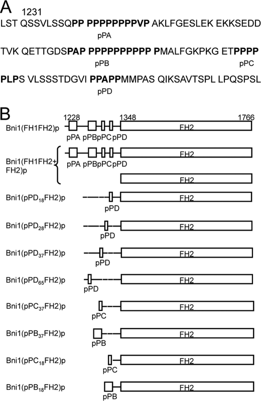
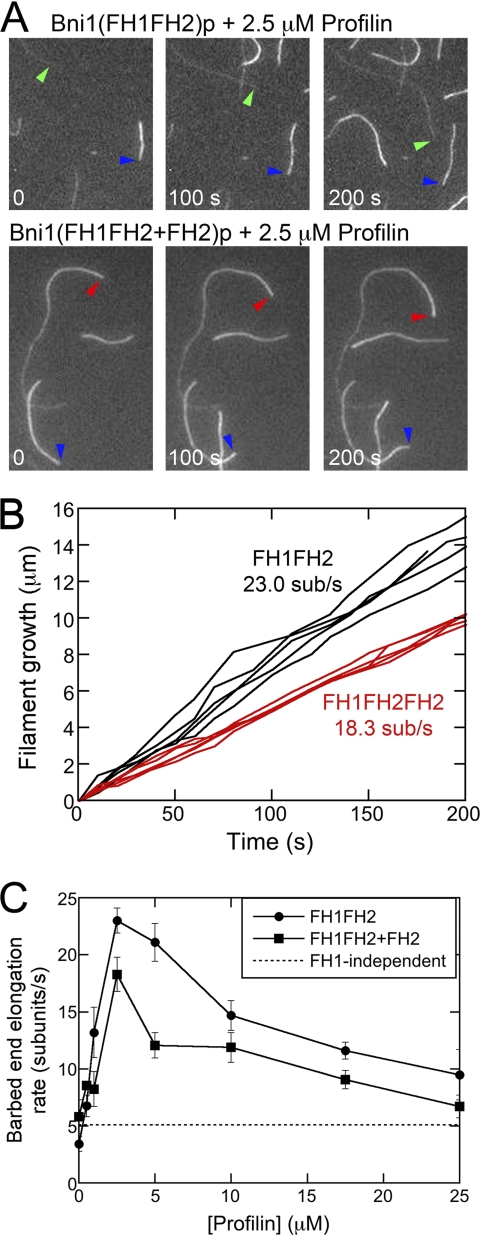
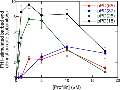
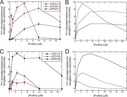
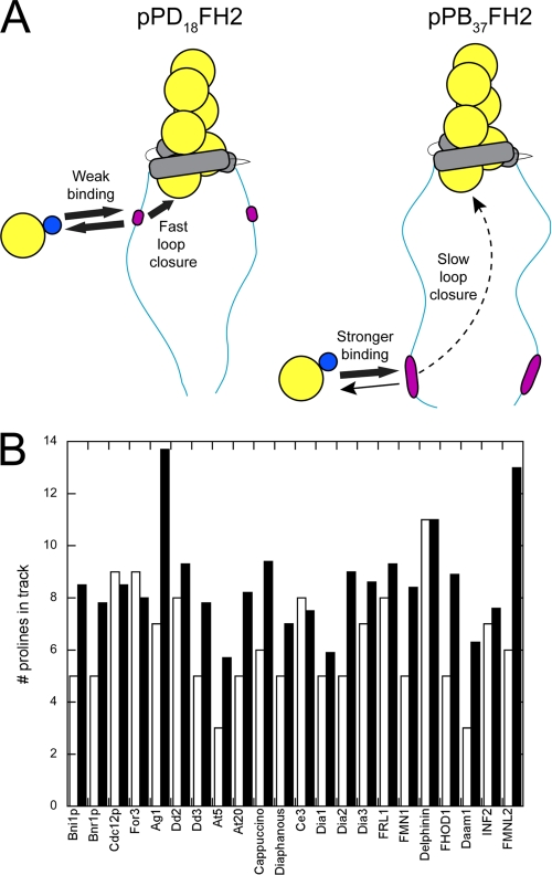
Formin-mediated elongation of actin filaments proceeds via association of Formin Homology 2 (FH2) domain dimers with the barbed end of the filament, allowing subunit addition while remaining processively attached to the end. The flexible Formin Homology 1 (FH1) domain, located directly N-terminal to the FH2 domain, contains one or more stretches of polyproline that bind the actin-binding protein profilin. Diffusion of FH1 domains brings associated profilin-actin complexes into contact with the FH2-bound barbed end of the filament, thereby enabling direct transfer of actin. We investigated how the organization of the FH1 domain of budding yeast formin Bni1p determines the rates of profilin-actin transfer onto the end of the filament. Each FH1 domain transfers actin to the barbed end independently of the other and structural evidence suggests a preference for actin delivery from each FH1 domain to the closest long-pitch helix of the filament. The transfer reaction is diffusion-limited and influenced by the affinities of the FH1 polyproline tracks for profilin. Position-specific sequence variations optimize the efficiency of FH1-stimulated polymerization by binding profilin weakly near the FH2 domain and binding profilin more strongly farther away. FH1 domains of many other formins follow this organizational trend. This particular sequence architecture may optimize the efficiency of FH1-stimulated elongation.


Figures

FIGURE 1.
FIGURE 1.
FH1 domain sequence and variants of Bni1(FH1FH2)p. A, sequence of FH1 domain. B, schematic representation of constructs used in this study. Residue numbers indicate domain boundaries. FH1 domains are to scale, but FH2 domains are not. All constructs form homodimers, except for the heterodimer of Bni1(FH1FH2+FH2)p. Both subunits are depicted in this case.
FIGURE 2.
FIGURE 2.
Effect of profilin on actin filament barbed end elongation mediated by Bni1p with a single FH1 domain. Conditions: 1.5 μm actin (33% Oregon Green) in microscopy buffer (9.6 mm imidazole (pH 7.0), 48 mm KCl, 0.96 mm MgCl2, 0.96 mm EGTA, 96 mm DTT, 1.92 mm ATP, 50 mm CaCl2, 14.4 mm glucose, 19.2 μg/ml catalase, 96 μg/ml glucose oxidase, 0.48% methylcellulose (4000 cP at 2%), 0.19% BSA). Data were collected with TIRFM. A and B, time series of images of formin-bound actin filaments growing in the presence of 20 nm wild-type Bni1(FH1FH2)p or 20 nm Bni1(FH1FH2 + FH2)p and 2.5 μm profilin. Colored arrows denote composition of barbed ends: blue marks free ends; green marks ends with Bni1(FH1FH2)p bound; and red marks ends with Bni1(FH1FH2+FH2)p bound. B, time courses of the growth of five filament barbed ends associated with Bni1(FH1FH2)p- (black data) or Bni1(FH1FH2+FH2)p- (red data) in the presence of 2.5 μm profilin. C, profilin concentration dependence of the elongation rates of barbed ends stimulated by wild-type Bni1(FH1FH2)p with two FH1 domains (circles) and one-armed Bni1(FH1FH2+FH2)p with one FH1 domain (squares). The dotted line represents the average contribution of FH1-independent polymerization, mediated through direct binding of actin subunits from the bulk phase, to the overall measured polymerization rates for wild-type and one-armed Bni1p. The error bars are S.E.
FIGURE 3.
FIGURE 3.
The effect of the distance between a single FH1 polyproline track pPD and the FH2 domain on Bni1p-mediated polymerization. Conditions: 1.5 μm actin (33% Oregon Green) in microscopy buffer with a range of profilin concentrations. Data were collected by TIRFM. Dependence of the FH1-stimulated barbed-end elongation rate of Bni1(pPD18FH2)p- (black), Bni1(pPD28FH2)p- (green), Bni1(pPD37FH2)p- (blue), and Bni1(pPD65FH2)p- (red) associated filaments on the concentration of profilin. The rate of FH2-mediated polymerization without profilin was subtracted from the polymerization rate measured at each profilin concentration to give the contribution of the pPD track to polymerization. The error bars are S.E.
FIGURE 4.
FIGURE 4.
Effect of sequence variations on FH1-stimulated polymerization. Conditions: 1.5 μm actin (33% Oregon Green) in microscopy buffer with varying concentrations of profilin. Data were collected by TIRFM. A, experimental and B, simulated dependence of FH1-stimulated barbed-end elongation on profilin concentration for variants of Bni1p with a single polyproline track (pPB, pPC, or pPD) located 37 residues (pPB position) from the FH2 domain. The error bars are S.E. C, experimental and D, simulated dependence of FH1-stimulated barbed-end elongation on profilin concentration for variants of Bni1p with a single polyproline track (pPB, pPC, or pPD) located 18 residues (pPD position) from the FH2 domain. The sequences inserted at these positions are the polyproline track sequences from sites pPB (red squares), pPC (blue diamonds), or pPD (black circles). Dissociation equilibrium constants for profilin:polyproline interactions are 20 μm for pPB, 600 μm for pPC, and 1200 μm for pPD. The loop closure rate for the profilin-bound FH1 domain is 10,000 s−1 at the pPB position and 50,000 s−1 at the pPD position. To account for a loss of FH1 flexibility upon insertion of the pPB sequence at the pPD position, the loop closure rate for free FH1 domain is 5,000 s−1 for pPB18FH2.
FIGURE 5.
FIGURE 5.
The position and length of polyproline tracks are tuned for efficient FH1-mediated polymerization. A, schematic representations of actin polymerization mediated by the pPD18FH2 versus pPB37FH2 constructs of Bni1(FH1FH2)p. Actin, profilin, FH2 domains, and polyproline tracks (pLp) are shown in yellow, blue, gray, and magenta. B, summary of the length of the polyproline track located closest to the FH2 domain (white bars) and the average length of polyproline tracks within an FH1 domain (black bars) for 22 formins. The formins listed are from S. cerevisiae (Bni1p and Bnr1p), S. pombe (Cdc12p and For3), A. gossypii (Ag1), D. discoideum (Dd2, Dd3), A. thaliana (At5, At20), D. melanogaster (Cappuccino, Diaphanous), C. elegans (Ce3), M. musculus (Dia1, Dia2, Dia3, FRL1, FMN1, Delphinin, FHOD1, Daam1), and H. sapiens (INF2, FMNL2).

References

    1. Chang F., Drubin D., Nurse P. (1997) cdc12p, a protein required for cytokinesis in fission yeast, is a component of the cell division ring and interacts with profilin. J. Cell Biol. 137, 169–182 - PMC - PubMed
    1. Watanabe N., Kato T., Fujita A., Ishizaki T., Narumiya S. (1999) Cooperation between mDia1 and ROCK in Rho-induced actin reorganization. Nat. Cell Biol. 1, 136–143 - PubMed
    1. Kobielak A., Pasolli H. A., Fuchs E. (2004) Mammalian formin-1 participates in adherens junctions and polymerization of linear actin cables. Nat. Cell Biol. 6, 21–30 - PMC - PubMed
    1. Pellegrin S., Mellor H. (2005) The Rho family GTPase Rif induces filopodia through mDia2. Curr. Biol.: CB 15, 129–133 - PubMed
    1. Pruyne D., Evangelista M., Yang C., Bi E., Zigmond S., Bretscher A., Boone C. (2002) Role of formins in actin assembly: nucleation and barbed-end association. Science 297, 612–615 - PubMed

Publication types

MeSH terms