

Case Reports
doi: 10.3348/kjr.2012.13.1.90.
Epub 2011 Dec 23.
Aggressive angiomyxoma: an unusual presentation
Affiliations
- PMID: 22247641
- PMCID: PMC3253408
- DOI: 10.3348/kjr.2012.13.1.90
Item in Clipboard
Case Reports
Aggressive angiomyxoma: an unusual presentation
Korean J Radiol.
2012 Jan-Feb.
Abstract
Aggressive angiomyxoma is an uncommon mesenchymal myxoid tumor that is characterized by slow growth and frequent local recurrence. It is currently regarded as a nonmetastasizing tumor. We describe a case of recurrent aggressive angiomyxoma with invasion into the veins including the inferior vena cava and the right atrium and with pulmonary metastases. Our case, together with those unusual cases documented in previous reports, may lead to a reappraisal of the nature of aggressive angiomyxoma.
Keywords: Aggressive angiomyxoma; Computed tomography; Histopathology; Inferior vena cava; Pulmonary metastases.
Figures
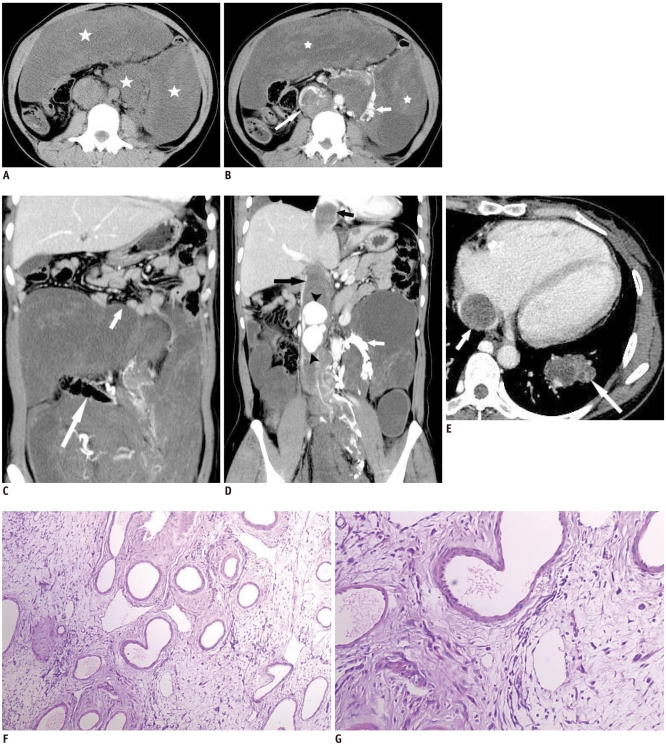
Aggressive angiomyxoma in 37-year-old woman. A. Unenhanced CT image shows large abdominal mass (stars) being hypo-attenuated in relation to surrounding muscular tissue. B. Contrast-enhanced CT image shows large homogeneous abdominal mass (stars). It directly invades inferior vena cava (long arrow), causing obstruction of inferior vena cava and formation of periaortic venous collaterals (short arrow). C. Coronal reconstruction image from contrast-enhanced CT shows mass encasing colon (long arrow) and causing portion of loop of small bowel to deviate superiorly (short arrow). D. Coronal reconstruction image from contrast-enhanced CT shows inferior vena cava filling defect (long black arrow) extending superiorly to level of right atrium (short black arrow). Image also shows prominent enhancing nodules within inferior vena cava (black arrowheads) and collateral vessels around aorta (white arrow). E. Contrast-enhanced CT image shows heterogeneously enhancing mass in left lower lobe (long arrow) and apparent inferior vena cava filling defect (short arrow). F. Low-power view shows vascular appearance of tumor, against myxoid, hypocellular background. G. Medium-power view shows bland cytological appearance of spindle cells.
References
-
- Sereda D, Sauthier P, Hadjeres R, Funaro D. Aggressive angiomyxoma of the vulva: a case report and review of the literature. J Low Genit Tract Dis. 2009;13:46–50. - PubMed
-
- Suleiman M, Duc C, Ritz S, Bieri S. Pelvic excision of large aggressive angiomyxoma in a woman: irradiation for recurrent disease. Int J Gynecol Cancer. 2006;16(Suppl 1):356–360. - PubMed
-
- Siassi RM, Papadopoulos T, Matzel KE. Metastasizing aggressive angiomyxoma. N Engl J Med. 1999;341:1772. - PubMed
-
- Blandamura S, Cruz J, Faure Vergara L, Machado Puerto I, Ninfo V. Aggressive angiomyxoma: a second case of metastasis with patient's death. Hum Pathol. 2003;34:1072–1074. - PubMed
-
- Steeper TA, Rosai J. Aggressive angiomyxoma of the female pelvis and perineum. Report of nine cases of a distinctive type of gynecologic soft-tissue neoplasm. Am J Surg Pathol. 1983;7:463–475. - PubMed
Publication types
MeSH terms
Substances
LinkOut - more resources
Full Text Sources
Medical
