. 2011 Dec;13(12):1101-12.
doi: 10.1593/neo.111060.

Merlin-deficient human tumors show loss of contact inhibition and activation of Wnt/β-catenin signaling linked to the PDGFR/Src and Rac/PAK pathways

Affiliations

Merlin-deficient human tumors show loss of contact inhibition and activation of Wnt/β-catenin signaling linked to the PDGFR/Src and Rac/PAK pathways

Lu Zhou et al. Neoplasia. 2011 Dec.

Abstract

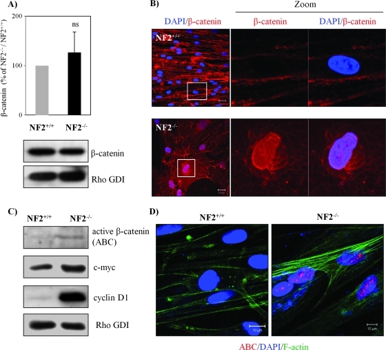
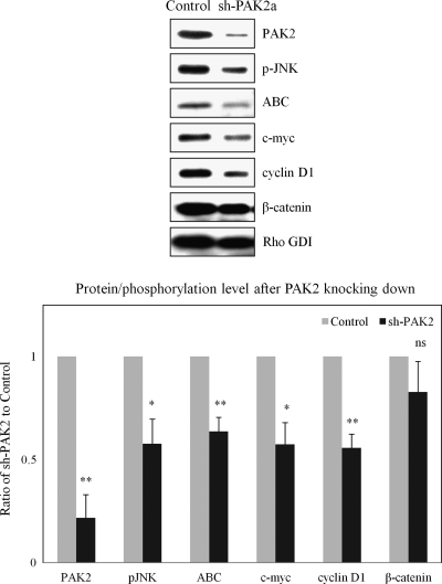
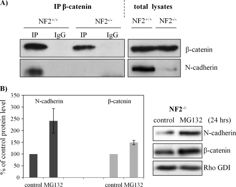
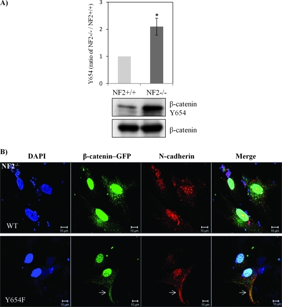
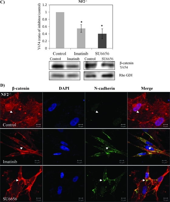
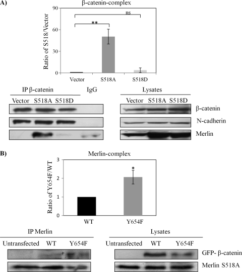
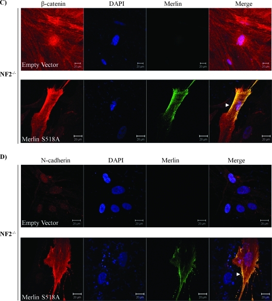
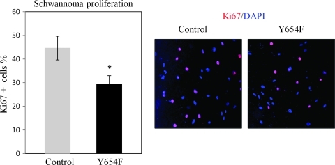
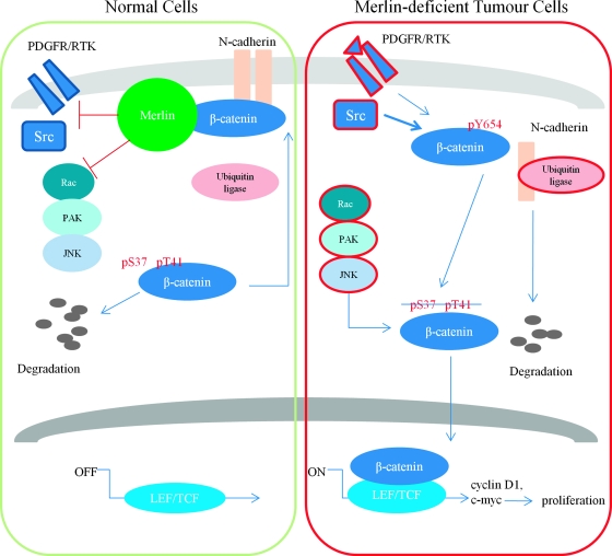
Neurofibromatosis type 2 (NF2) is an inherited predisposition cancer syndrome characterized by the development of multiple benign tumors in the nervous system including schwannomas, meningiomas, and ependymomas. Using a disease model comprising primary human schwannoma cells, we previously demonstrated that adherens junctions (AJs) are impaired in schwannoma cells because of a ubiquitous, upregulated Rac activity. However, the mechanism by which loss of contact inhibition leads to proliferation remains obscure in merlin-deficient tumors. In this study, we show that proliferative Wnt/β-catenin signaling is elevated as active β-catenin (dephosphorylated at serine 37 and threoine 41) localizes to the nucleus and the Wnt targets genes c-myc and cyclin D1 are upregulated in confluent human schwannoma cells. We demonstrate that Rac effector p21-activated kinase 2 (PAK2) is essential for the activation of Wnt/β-catenin signaling because depletion of PAK2 suppressed active β-catenin, c-myc, and cyclin D1. Most importantly, the link between the loss of the AJ complex and the increased proliferation in human schwannoma cells is connected by Src and platelet-derived growth factor receptor-induced tyrosine 654 phosphorylation on β-catenin and associated with degradation of N-cadherin. We also demonstrate that active merlin maintains β-catenin and N-cadherin complex at the plasma membrane through direct regulation. Finally, we demonstrate that phosphorylation of tyrosine 654 is critical for the increased proliferation in human schwannoma cells because overexpression of a Y654F mutant β-catenin reduces hyperproliferation of schwannoma cells. We suggest a model that these pathways are coordinated and relevant for proliferation in merlin-deficient tumors.


Figures

Figure 1
Figure 1
Wnt signaling is elevated in human schwannoma cells. (A) Western blot analyses were carried out to compare the levels of β-catenin between Schwann and schwannoma cells. RhoGDI was used as a loading control between Schwann and schwannoma cells. No significant difference (ns) was observed after quantification and statistical analysis (n = 5). Error bars represent the mean ± SEM. (B) β-Catenin localizes in the nucleus of schwannoma but not Schwann cells. Confluent and 24-hour starved human Schwann (NF2+/+) and schwannoma cells (NF2-/-) were immunostained with DAPI (nuclear marker) and β-catenin. Scale bar, 20 µm. (C) ABC and Wnt target genes are elevated in schwannoma. Western blot analyses were carried out to compare the level of ABC, c-myc, and cyclin D1. (D) The nuclear portion of β-catenin in schwannoma is ABC. Immunostaining was carried out for ABC, DAPI, and cytoskeleton marker F-actin in Schwann and schwannoma cells. Scale bar, 10 µm.
Figure 2
Figure 2
Knockdown PAK2 suppresses Wnt/β-catenin signaling. Schwannoma cells were infected with shRNA encoding a sequence targeting human PAK2 (sh-PAK2a) or a matched scramble sequence (Control). Western blot analyses were carried out to compare the levels of pJNK, ABC, c-myc, cyclin D1, and β-catenin between sh-PAK2a and control. Densities of bands were quantified and compared. Error bars represent the mean ± SEM. n = 3. *P < .05. **P < .01. ns indicates no significant difference. pJNK, ABC, c-myc, and cyclin D1 were significantly reduced after knocking down PAK2.
Figure 3
Figure 3
N-cadherin reduced in schwannoma cells and its degradation. (A) Schwann (NF2+/+) and schwannoma cells (NF2-/-) were lysed in NP40 buffer and immunoprecipitated with an antibody against β-catenin. IgG mouse without anti-β-catenin antibody served as control. Western blot analysis was carried out to detect the levels of β-catenin and N-cadherin in both co-IP complex and total lysates. (B) N-cadherin is accumulated after MG132 inhibition. Schwannoma cells were treated with MG132 (1 µM) for 24 hours, Western blots were then carried out for N-cadherin and β-catenin. RhoGDI served as loading control. Error bars represent the mean ± SEM.
Figure 4
Figure 4
β-Catenin Y654 phosphorylation is enhanced in schwannoma cells and is mediated by Src and PDGFR at the plasma membrane. (A) β-Catenin Y654 phosphorylation is elevated in schwannomas. Confluent Schwann cells (NF2+/+) and schwannoma cells (NF2-/-) was starved for 24 hours before being treated with pervanadate for 30 minutes to reserve the phosphorylation signals. Western blot analysis was carried out to compare the level of phosphorylation of β-catenin at tyrosine 654. Total β-catenin served as control. Densities of bands were quantified and compared. (B) β-Catenin Y654F relocates N-cadherin to the membrane in schwannoma cells (NF2-/-). Cells were transfected with GFP-β-catenin-WT and GFP-β-catenin-Y654F, respectively. Immunostaining was carried out with DAPI, anti-GFP, and anti-N-cadherin. Scale bar, 10 µm. The sites at the plasma membrane are indicated by arrows. (C) PDGFR and Src inhibitors (imatinib and SU6656) reduced β-catenin Y654 phosphorylation. Schwannoma cells were starved for 24 hours and then treated with DMSO or different inhibitors as well as pervanadate for 30 minutes. Western blot analyses were carried out for β-catenin Y654. RhoGDI was used as a loading control. Densities of bands were quantified and compared. (D) PDGFR and Src inhibitors bring the colocalization of N-cadherin and β-catenin back to the plasma membrane. Cells were treated with control (DMSO), imatinib, and SU6656 for 24 hours. Immunostaining was then carried out with DAPI, anti-N-cadherin, and anti-β-catenin. The potential AJ sites are indicated by arrows. Scale bar, 10 µm.
Figure 4
Figure 4
β-Catenin Y654 phosphorylation is enhanced in schwannoma cells and is mediated by Src and PDGFR at the plasma membrane. (A) β-Catenin Y654 phosphorylation is elevated in schwannomas. Confluent Schwann cells (NF2+/+) and schwannoma cells (NF2-/-) was starved for 24 hours before being treated with pervanadate for 30 minutes to reserve the phosphorylation signals. Western blot analysis was carried out to compare the level of phosphorylation of β-catenin at tyrosine 654. Total β-catenin served as control. Densities of bands were quantified and compared. (B) β-Catenin Y654F relocates N-cadherin to the membrane in schwannoma cells (NF2-/-). Cells were transfected with GFP-β-catenin-WT and GFP-β-catenin-Y654F, respectively. Immunostaining was carried out with DAPI, anti-GFP, and anti-N-cadherin. Scale bar, 10 µm. The sites at the plasma membrane are indicated by arrows. (C) PDGFR and Src inhibitors (imatinib and SU6656) reduced β-catenin Y654 phosphorylation. Schwannoma cells were starved for 24 hours and then treated with DMSO or different inhibitors as well as pervanadate for 30 minutes. Western blot analyses were carried out for β-catenin Y654. RhoGDI was used as a loading control. Densities of bands were quantified and compared. (D) PDGFR and Src inhibitors bring the colocalization of N-cadherin and β-catenin back to the plasma membrane. Cells were treated with control (DMSO), imatinib, and SU6656 for 24 hours. Immunostaining was then carried out with DAPI, anti-N-cadherin, and anti-β-catenin. The potential AJ sites are indicated by arrows. Scale bar, 10 µm.
Figure 5
Figure 5
Only Merlin S518A forms a complex with β-catenin and relocates AJs complex to the plasma membrane. (A) β-Catenin forms a complex with Merlin-S518A not Merlin S518D. HEK293T cells are transfected with empty vector, Merlin-S518A, and Merlin-S518D. Endogenous β-catenin complex was immunoprecipitated (IP) after treatment with pervanadate to reserve the phosphorylation status of β-catenin. IgG mouse without anti-β-catenin antibody served as a control. Immunoprecipitates were blotted for β-catenin. Merlin, N-cadherin, and RhoGDI served as controls. Densities of bands were quantified and compared. The protein level of merlin in the β-catenin complex was compared between Merlin S518 mutants and empty vector after being corrected with their count parts in total lysates. Error bars represent the mean ± SEM. n = 3. **P < .01. ns indicates no significant difference. (B) Merlin-S518A preferentially interacts with β-catenin-Y654F. Merlin-S518A stable line (HEK293T cells) was transfected with GFP-β-catenin WT and GFP-β-catenin-Y654F (Y654F), and untransfected cells. Merlin complex was immunoprecipitated and blotted for β-catenin and merlin. Densities of bands were quantified and compared. The protein level of β-catenin in the merlin complex was compared between WT and Y654F after being corrected with their count parts in total lysates. Error bars represent the mean ± SEM. n = 3. *P < .05. (C, D) Merlin-S518A relocates β-catenin and N-cadherin to the plasma membrane in schwannoma cells. Cells were transfected with either empty vector or Merlin-S518A. Immunostaining was carried out for merlin and β-catenin (C) or merlin and N-cadherin (D). DAPI was used as nuclear maker. The potential AJ sites are indicated by arrows. Scale bar, 20 µm.
Figure 5
Figure 5
Only Merlin S518A forms a complex with β-catenin and relocates AJs complex to the plasma membrane. (A) β-Catenin forms a complex with Merlin-S518A not Merlin S518D. HEK293T cells are transfected with empty vector, Merlin-S518A, and Merlin-S518D. Endogenous β-catenin complex was immunoprecipitated (IP) after treatment with pervanadate to reserve the phosphorylation status of β-catenin. IgG mouse without anti-β-catenin antibody served as a control. Immunoprecipitates were blotted for β-catenin. Merlin, N-cadherin, and RhoGDI served as controls. Densities of bands were quantified and compared. The protein level of merlin in the β-catenin complex was compared between Merlin S518 mutants and empty vector after being corrected with their count parts in total lysates. Error bars represent the mean ± SEM. n = 3. **P < .01. ns indicates no significant difference. (B) Merlin-S518A preferentially interacts with β-catenin-Y654F. Merlin-S518A stable line (HEK293T cells) was transfected with GFP-β-catenin WT and GFP-β-catenin-Y654F (Y654F), and untransfected cells. Merlin complex was immunoprecipitated and blotted for β-catenin and merlin. Densities of bands were quantified and compared. The protein level of β-catenin in the merlin complex was compared between WT and Y654F after being corrected with their count parts in total lysates. Error bars represent the mean ± SEM. n = 3. *P < .05. (C, D) Merlin-S518A relocates β-catenin and N-cadherin to the plasma membrane in schwannoma cells. Cells were transfected with either empty vector or Merlin-S518A. Immunostaining was carried out for merlin and β-catenin (C) or merlin and N-cadherin (D). DAPI was used as nuclear maker. The potential AJ sites are indicated by arrows. Scale bar, 20 µm.
Figure 6
Figure 6
Overexpression of β-catenin Y654F inhibits the hyperproliferation of human schwannoma cells. Cells were transfected with GFP-β-catenin-Y654F (Y654F) and empty vector (control), starved for 24 hours, and stimulated with GFM for another 24 hours and then stained with DAPI and Ki67. The stained cells were counted manually; the Ki67-positive (ratio of Ki67 to DAPI) cells were calculated. Error bars represent the mean ± SEM. n = 3. *P < .05, t test.
Figure 7
Figure 7
Hypothetical model of merlin's role on inhibition of Wnt/β-catenin signaling. In normal cells, merlin inhibits Src/PDGFR, Rac/PAK/JNK, and maybe N-cadherin ubiquitination as well to keep Y654 unphosphorylated β-catenin staying at AJ together with N-cadherin. In addition, excess cytoplasmic β-catenin can be regulated by proteasome degradation. In merlin-deficient tumor cells, loss of merlin leads to activation of PDGFR and Src (red). β-Catenin is then phosphorylated by Src/PDGFR at Y654 and disassociated with N-cadherin. In further regulation by activated Rac/PAK/JNK (red), β-catenin shuttles into the nucleus to activate transcriptional factors LEF/TCF and then drive the expression of downstream targets, namely, cyclin D1, c-myc, and eventually, proliferation is increased in tumor cells.

References

    1. Hanemann CO. Magic but treatable? Tumours due to loss of merlin. Brain. 2008;131:606–615. - PubMed
    1. McClatchey AI, Fehon RG. Merlin and the ERM proteins—regulators of receptor distribution and signaling at the cell cortex. Trends Cell Biol. 2009;19:198–206. - PMC - PubMed
    1. Li W, You L, Cooper J, Schiavon G, Pepe-Caprio A, Zhou L, Ishii R, Giovannini M, Hanemann CO, Long SB, et al. Merlin/NF2 suppresses tumorigenesis by inhibiting the E3 ubiquitin ligase CRL4(DCAF1) in the nucleus. Cell. 2010;140:477–490. - PMC - PubMed
    1. Ammoun S, Flaiz C, Ristic N, Schuldt J, Hanemann CO. Dissecting and targeting the growth factor-dependent and growth factor-independent extracellular signal-regulated kinase pathway in human schwannoma. Cancer Res. 2008;68:5236–5245. - PubMed
    1. Curto M, Cole BK, Lallemand D, Liu CH, McClatchey AI. Contact-dependent inhibition of EGFR signaling by Nf2/Merlin. J Cell Biol. 2007;177:893–903. - PMC - PubMed

Publication types

MeSH terms

LinkOut - more resources