. 2012;7(1):e28626.
doi: 10.1371/journal.pone.0028626. Epub 2012 Jan 11.

Anti-proteinase 3 anti-neutrophil cytoplasm autoantibodies recapitulate systemic vasculitis in mice with a humanized immune system

Affiliations

Anti-proteinase 3 anti-neutrophil cytoplasm autoantibodies recapitulate systemic vasculitis in mice with a humanized immune system

Mark A Little et al. PLoS One. 2012.

Abstract

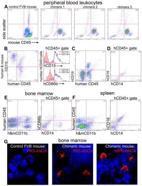
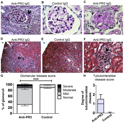
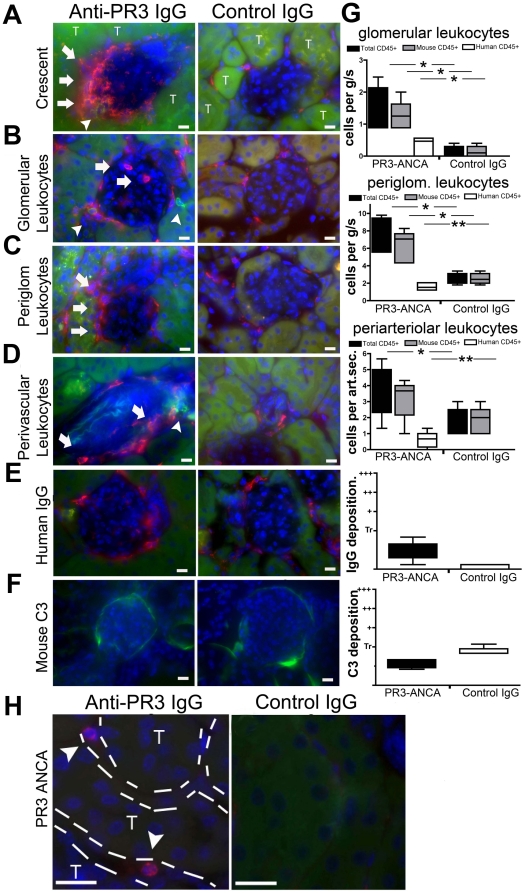
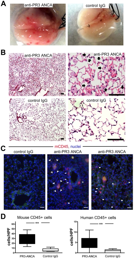
Evidence is lacking for direct pathogenicity of human anti-proteinase-3 (PR3) antibodies in development of systemic vasculitis and granulomatosis with polyangiitis (GPA, Wegener's granulomatosis). Progress in study of these antibodies in rodents has been hampered by lack of PR3 expression on murine neutrophils, and by different Fc-receptor affinities for IgG across species. Therefore, we tested whether human anti-PR3 antibodies can induce acute vasculitis in mice with a human immune system. Chimeric mice were generated by injecting human haematopoietic stem cells into irradiated NOD-scid-IL2Rγ⁻/⁻ mice. Matched chimera mice were treated with human IgG from patients with: anti-PR3 positive renal and lung vasculitis; patients with non-vasculitic renal disease; or healthy controls. Six-days later, 39% of anti-PR3 treated mice had haematuria, compared with none of controls. There was punctate bleeding on the surface of lungs of anti-PR3 treated animals, with histological evidence of vasculitis and haemorrhage. Anti-PR3 treated mice had mild pauci-immune proliferative glomerulonephritis, with infiltration of human and mouse leukocytes. In 3 mice (17%) more severe glomerular injury was present. There were no glomerular changes in controls. Human IgG from patients with anti-PR3 autoantibodies is therefore pathogenic. This model of anti-PR3 antibody-mediated vasculitis may be useful in dissecting mechanisms of microvascular injury.


Conflict of interest statement

Competing Interests: C.O.S. is employed by GSK. This study was partly funded by a GRIP award from Genzyme. There are no patents, products in development or marketed products to declare. This does not alter the authors' adherence to all the PLoS ONE policies on sharing data and materials.

Figures

Figure 1
Figure 1. Characterization of chimerism in NOD-scid-IL2Rγ−/− mice.
(A–D) Flow cytometric analysis of leukocytes from tail bleeds six weeks after administration of HSCs (n = 26 mice). (A) Plots showing mouse leukocytes labelled with anti-mouse CD45 antibodies. Compared with control wild-type mouse blood, chimeras have populations of mCD45 negative leukocytes that show SSC characteristics of granulocytes (High), monocytes (Int) and lymphocytes (low). (B) Chimera blood leukocytes express human CD45 and many of these express CD11b. hCD45+,CD11b+ leukocytes predominantly express hCD15 and hCD66b compared with hCD45+,CD11b− leukocytes shown in histograms. (C) A proportion of hCD45+ leukocytes express CD19. (D) Some hCD45 leukocytes are CD14high and some are CD16+,CD14low. (E) In chimera bone marrow there are CD11b+ leukocytes which do not express mCD45 and among hCD45+ leukocytes a proportion express CD14 and a proportion express CD66b. (F) In chimera spleen there are CD11b+ leukocytes which express hCD45 and among hCD45+ leukocytes many express both CD14 and CD16. (G) Bone marrow spreads from wild type or chimera mice, labelled with anti-hMPO or anti-hPR3 IgG antibodies (red) purified from patients with vasculitis. Note that chimera bone marrow demonstrates anti-hMPO or anti-hPR3 antibody positive leukocytes with characteristic human neutrophil nuclear morphology. Wild type mouse bone marrow shows no cells positive for these antigens indicating that the anti-human antibodies do not cross react with mouse neutrophils.
Figure 2
Figure 2. Anti-PR3 antibodies cause kidney disease.
(A–C) PAS stained images of glomeruli from chimera mice 6 days after injection with anti-PR3 (n = 18, A, 400×; C, 600×) or control IgG (n = 8, B, 600×). Note extra-capillary proliferation and peri-glomerular inflammation (arrowhead) (A), and mesangiolysis (C, arrow) in anti-PR3 treated mice. (D–F) H & E stained sections of kidney from chimera mice treated with anti-PR3 (D, 40×) or disease control (E, 40×) IgG. There are regions of tubulointerstitial injury, with red cell cast formation (arrow). (F) Demonstrates intense peri-glomerular inflammation in an animal treated with anti-PR3 IgG (arrowhead, 400×). By comparison mice treated with disease control IgG showed minimal glomerular or tubulointerstitial changes. (G) Fractions of glomeruli affected in anti-PR3 (n = 18) and control IgG (n = 8) treated animals (Error bars depict SEM; ***p = 0.001) (H). Degree of tubulointerstitial disease in mice treated with anti-PR3 antibodies and control antibodies (*P<0.05, median ± IQ ± max/min values). (Bars = 50 µm).
Figure 3
Figure 3. Anti-PR3 antibodies induce infiltration of kidneys with leukocytes of murine and human origin.
Kidney sections were incubated with anti-mCD45 (red) and anti-hCD45 (green) antibodies and images were captured by fluorescence microscopy (T = tubule). Occasional (<5%) glomeruli of anti-PR3 treated mice displayed intense extracapillary leukocyte infiltration (A) in the shape of crescents (arrows). Most glomeruli in animals treated with anti-PR3 antibodies (n = 18) had evidence of intraglomerular (B,G) and peri-glomerular (C,G) leukocyte infiltration. These were comprised mostly of mCD45+ cells, although some hCD45 leukocytes were also present (arrowheads). In addition, there was a significant increase in peri-vascular leukocyte (mCD45+ and hCD45+) infiltration in anti-PR3 treated mice (D,G [per arteriolar section (art.sec.)]). Sections were also stained for deposition of IgG [red] (E,G) and C3 [green] (F,G). IgG was detectable within periglomerular cells, but there was minimal deposition within the glomeruli. Mouse C3 was weakly deposited in glomeruli but was no different between control group (n = 8) and anti-PR3 group (n = 18). Note mouse C3 can be detected normally binding avidly to tubular basement membranes. (Marker = 10 µm) (*P<0.05, **P<0.01. median ± IQ ± max/min values). (H) Kidney sections from anti-PR3 and control treated animals were incubated with anti-PR3 positive ANCA IgG. In the peritubular capillaries of chimera mice that received anti-PR3 hIgG occasional leukocytes detected by anti-hPR3 hIgG could be detected. No positively stained human neutrophils were seen in glomeruli.
Figure 4
Figure 4. Anti-PR3 antibodies induce capillaritis with leukocytes of mouse and human origin.
(A) Photomicrographs of explanted lungs from mice treated with disease control IgG (n = 8) or from patients with anti-PR3 ANCA (n = 18). Mice treated with anti-PR3 ANCA displayed petechiae over the surface of the lung. (B) H&E stained low and high power images of lungs from chimera mice injected with anti-PR3 antibodies 6 days previously. Note hemorrhage, inflammation, thickening of alveolar walls, enlarged, highly vacuolated alveolar macrophages and prominent recruitment of neutrophils (arrows) within the alveolar walls. These features are consistent with capillaritis. By comparison chimera mice treated with control IgG showed normal lung architecture. (C) Immunostaining of lung tissue to assess the degree of leukocyte infiltration. Note that in mice that received anti-PR3 ANCA IgG there was significant recruitment of leukocytes to the peribronchial areas (thin arrows) and also the alveolar areas (fat arrows). In addition many alveoli in mice treated with anti-PR3 antibodies had apoptotic debris that was partially ingested by alveolar macrophages (arrowheads) (bar = 50 µm). (D) Blinded assessment of human and mouse leukocyte recruitment to the lungs of treated animals (** P<0.01. median ± IQ ± max/min values).

References

    1. van der Woude FJ, Rasmussen N, Lobatto S, Wiik A, Permin H, et al. Autoantibodies against neutrophils and monocytes: tool for diagnosis and marker of disease activity in Wegener's granulomatosis. Lancet. 1985;1(8426):425–9. - PubMed
    1. Falk RJ, Jennette JC. Anti-neutrophil cytoplasmic autoantibodies with specificity for myeloperoxidase in patients with systemic vasculitis and idiopathic necrotizing and crescentic glomerulonephritis. N Engl J Med. 1988;318:1651–7. - PubMed
    1. Niles JL, McCluskey RT, Ahmad MF, Arnaout MA. Wegener's granulomatosis autoantigen is a novel neutrophil serine proteinase. Blood. 1989;74:1888–93. - PubMed
    1. Jennette JC, Hoidal JR, Falk RJ. Specificity of anti-neutrophil cytoplasmic autoantibodies for proteinase 3. Blood. 1990;75:2263–4. - PubMed
    1. Goldschmeding R, van der Schoot CE, ten Bokkel Huinink D, Hack CE, van den Ende ME, et al. Wegener's granulomatosis autoantibodies identify a novel diisopropylfluorophosphate-binding protein in the lysosomes of normal human neutrophils. J Clin Invest. 1989;84:1577–87. - PMC - PubMed

Publication types

MeSH terms