. 2012;7(1):e29608.
doi: 10.1371/journal.pone.0029608. Epub 2012 Jan 11.

The organisation of Ebola virus reveals a capacity for extensive, modular polyploidy

Affiliations

The organisation of Ebola virus reveals a capacity for extensive, modular polyploidy

Daniel R Beniac et al. PLoS One. 2012.

Abstract

Background: Filoviruses, including Ebola virus, are unusual in being filamentous animal viruses. Structural data on the arrangement, stoichiometry and organisation of the component molecules of filoviruses has until now been lacking, partially due to the need to work under level 4 biological containment. The present study provides unique insights into the structure of this deadly pathogen.

Methodology and principal findings: We have investigated the structure of Ebola virus using a combination of cryo-electron microscopy, cryo-electron tomography, sub-tomogram averaging, and single particle image processing. Here we report the three-dimensional structure and architecture of Ebola virus and establish that multiple copies of the RNA genome can be packaged to produce polyploid virus particles, through an extreme degree of length polymorphism. We show that the helical Ebola virus inner nucleocapsid containing RNA and nucleoprotein is stabilized by an outer layer of VP24-VP35 bridges. Elucidation of the structure of the membrane-associated glycoprotein in its native state indicates that the putative receptor-binding site is occluded within the molecule, while a major neutralizing epitope is exposed on its surface proximal to the viral envelope. The matrix protein VP40 forms a regular lattice within the envelope, although its contacts with the nucleocapsid are irregular.

Conclusions: The results of this study demonstrate a modular organization in Ebola virus that accommodates a well-ordered, symmetrical nucleocapsid within a flexible, tubular membrane envelope.


Conflict of interest statement

Competing Interests: The authors have declared that no competing interests exist.

Figures

Figure 1
Figure 1. Quantitation of Ebola virus length.
(A) Histogram of virion length, with cryo-EM images showing single, continuous and linked particles. A total of 2090 virions with continuous nucleocapsids (no obvious segmentations) were measured, showing the relationship between length and genome copy number per virus. Empty and linked EBOV structures were excluded from the histogram data. A single G1-single/comma shaped EBOV is shown (inset on the right, G1 = 1 copy of genome). (B) Low magnification cryo-images showing: G1- single/comma shape, G1- single/linear, G5-continuous (G5 = 5 copies of genome). (C) High magnification of a G1- (single genome) virion with a region filtered to emphasize the nucleocapsid. (D) Low magnification image of a G4-linked EBOV, each genome copy is indicated and numbered, the red arrows show the transition points between nucleocapsids. The circular holes (filled with vitreous ice) appear as lighter regions and the support film (“quantifoil”) appears dark grey. A “linker” region is shown at higher magnification (inset).
Figure 2
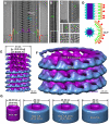
Figure 2. Image processing of Ebola virus.
Linear 2D averaging of EBOV: the envelope and nucleocapsid are prominent features (A). The line trace is colour-coded as follows: red, spike; beige, lipid envelope; green, membrane-associated proteins; white, membrane-nucleocapsid gap; blue and purple, outer and inner nucleocapsid. (B) 2D class averages of envelope plus inner face. (C) VP40 VLPs, showing 2D averages from the from side regions (first two) and end-on/central regions (last three). In (A–C) representative individual repeats have been highlighted in color using the same scheme as in (A). (D) Schematic model of the nucleocapsid and envelope, highlighting the relative distribution of NP to VP40. (E,F) 3D reconstruction of the nucleocapsid with the same colour scheme as in (A). The location of the inner nucleocapsid, and the bridge are indicated. The reconstruction is presented at a volume threshold that would encompass a single copy of each of these proteins, and the viral RNA. In (E) the vertical (protein-protein) and horizontal (protein-RNA) contacts are indicated by yellow and white arrows, respectively. (G) Various recombinant nucleocapsid-like structures, and authentic EBOV, which have been studied by electron microscopy , , , . 3D schematics of these structures highlighting the RNA and protein composition and the diameter of these structures, at the same scale for comparison to (E).
Figure 3
Figure 3. Electron tomography of Ebola virus.
(A–C) Cryo-EM images of; (A) VP40-GP VLP, (B) VP40 VLP, and (C) EBOV. (D–F) Electron tomogram of VP40-GP VLP showing; (D) a single X–Y slice cutting through the spikes at the top of the VLP, (E) a single X–Y slice cutting through the central region of the VLP, and (F) a X–Z average of 50 slices showing a cross section of the VLP. (G–H) Electron tomograms of EBOV showing; (G) a single X–Y slice cutting through the central region of the virus, and (H) a X–Z average of 50 slices showing a cross section of the virus. The insets in (G) are image averages of the nucleocapsid (bottom), and the entire width of the virus (top). (I) 3D shaded surface representation of the EBOV tomogram, individual spikes (red oval) and connective regions between the nucleocapsid and membrane proteins (red rectangle) have been highlighted. (J) Surface distribution of the spikes on the VP40-GP VLP tomogram shown in (D–F), the envelope has been replaced with an orange cylinder, and spike locations are indicated by white spheres. (K) 3D model of EBOV using the data from Figs. 2, 3, 4. Color coding as follows: red, spike; white spheres, spike location; beige/orange, lipid envelope; green, membrane associated proteins; blue and purple, outer and inner nucleocapsid. Colour-coded arrows in (A–H) highlight equivalent features in the 2D analysis shown in Fig. 2.
Figure 4
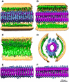
Figure 4. Sub-tomogram averaging of Ebola virus.
(A–D) Sections of the density map of the sub-tomogram average are shown from the top sliced just below the envelope (A), the middle of the virus (B), a side view of the virus (C), and an end-on slice (D). Putative locations of several VP40 proteins adjacent to the membrane are circled. (E,F) Images showing just the nucleocapsid. The helix is right handed (arrow in E). No helical symmetry was applied to this data. Color coding as follows; beige, lipid envelope; green, membrane associated proteins (VP40); blue and purple, outer and inner nucleocapsid.
Figure 5
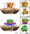
Figure 5. 3D structure of the Ebola spike.
The density map of the EBOV GP spike viewed from the side, end-on, and side (with envelope) shows the docked GP1–GP2 structure in yellow (PDB entry 3CSY [42]), glycosylation sites (green), and receptor binding site (RBS; red, highlighted). (A) The reconstruction showing the spike (orange) and the envelope (beige). (B) Difference map generated by subtracting the docked structure from reconstruction of the entire spike. The color scheme shows the following putative regions; green, mucin domain; pink, deletions 190–213, 279–298; purple-blue, GP2 stalk. The docked KZ52 neutralizing antibody is shown in purple.
Figure 6
Figure 6. Schematic model of Ebola virus genome packaging.
EBOV appears pleomorphic, but an underlying structural organization is maintained. In the model we show the three basic morphological forms of EBOV particles; empty, linked, and continuous. Single genome (G1) virus and multi-genome particles are shown budding from the cell. In this model genomes are assembled in the host cell and transported to the surface where the end-to-end apposition that we have observed by cryo-EM in mature virions takes place during (or prior to) budding and envelopment at the plasma membrane. The color-coding is as follows: nucleocapsids, red, yellow and orange helices; nucleocapsid protein, purple spheres; VP40, green ovals, VP24/VP35 bridges, blue oval; GP spikes, red; microtubules, brown.

References

    1. Caspar DL, Klug A. Physical principles in the construction of regular viruses. Cold Spring Harb Symp Quant Biol. 1962;27:1–24. - PubMed
    1. Hosaka Y, Kitano H, Ikeguchi S. Studies on the pleomorphism of HVJ virons. Virology. 1966;29:205–221. - PubMed
    1. Luque D, Rivas G, Alfonso C, Carrascosa JL, Rodriguez JF, et al. Infectious bursal disease virus is an icosahedral polyploid dsRNA virus. Proc Natl Acad Sci U S A. 2009;106:2148–2152. - PMC - PubMed
    1. Noda T, Sagara H, Yen A, Takada A, Kida H, et al. Architecture of ribonucleoprotein complexes in influenza A virus particles. Nature. 2006;439:490–492. - PubMed
    1. Geisbert TW, Hensley LE. Ebola virus: new insights into disease aetiopathology and possible therapeutic interventions. Expert Rev Mol Med. 2004;6:1–24. - PubMed

Publication types