A case of portal vein thrombosis by protein C and s deficiency completely recanalized by anticoagulation therapy
- PMID: 22247922
- PMCID: PMC3252510
- DOI: 10.4068/cmj.2011.47.3.185
A case of portal vein thrombosis by protein C and s deficiency completely recanalized by anticoagulation therapy
Abstract
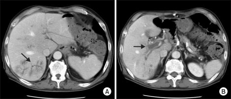
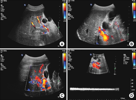
Portal vein thrombosis (PVT) is a rare form of venous thrombosis that affects the hepatic portal vein flow, which can lead to portal hypertension. Treatment of PVT includes anticoagulants, thrombolysis, insertion of shunts, bypass surgery, and liver transplantation. Single anticoagulation therapy is not regarded as a curative treatment but can be associated with a reduction in new thrombotic episodes. We experienced a case of acute total occlusion of PVT provoked by protein C and S deficiency syndrome. PVT was completely recanalized with oral anticoagulant therapy following low molecular weight heparin therapy.
Keywords: Anticoagulation; Protal vein; Protein C deficiency; Protein S deficiency; Thrombosis.
Figures

Similar articles
-
Portal vein thrombosis and liver cirrhosis: Long-term anticoagulation is effective and safe.Clin Res Hepatol Gastroenterol. 2019 Aug;43(4):395-402. doi: 10.1016/j.clinre.2018.11.011. Epub 2018 Dec 18. Clin Res Hepatol Gastroenterol. 2019. PMID: 30578107
-
Portal vein thrombosis after laparoscopic sleeve gastrectomy: presentation and management.Surg Obes Relat Dis. 2016 Dec;12(10):1787-1794. doi: 10.1016/j.soard.2016.03.005. Epub 2016 Mar 8. Surg Obes Relat Dis. 2016. PMID: 27178606
-
Portal vein thrombosis with protein C-S deficiency in a non-cirrhotic patient.World J Hepatol. 2014 Jul 27;6(7):532-7. doi: 10.4254/wjh.v6.i7.532. World J Hepatol. 2014. PMID: 25068006 Free PMC article.
-
Nontumoral Portal Vein Thrombosis: A Challenging Consequence of Liver Cirrhosis.J Clin Transl Hepatol. 2020 Dec 28;8(4):432-444. doi: 10.14218/JCTH.2020.00067. Epub 2020 Nov 11. J Clin Transl Hepatol. 2020. PMID: 33447527 Free PMC article. Review.
-
Portal vein thrombosis in cirrhosis.J Clin Exp Hepatol. 2014 Dec;4(4):320-31. doi: 10.1016/j.jceh.2013.12.003. Epub 2013 Dec 31. J Clin Exp Hepatol. 2014. PMID: 25755579 Free PMC article. Review.
Cited by
-
Multiple abdominal veins thrombosis secondary to protein s deficiency - a case report.J Clin Diagn Res. 2014 Jun;8(6):MD07-8. doi: 10.7860/JCDR/2014/5879.4509. Epub 2014 Jun 20. J Clin Diagn Res. 2014. PMID: 25121018 Free PMC article.
-
Recanalization of port-superior mesenteric vein thrombosis with long-term anticoagulant therapy after failed early anticoagulant therapy.Surg Case Rep. 2024 Jun 20;10(1):154. doi: 10.1186/s40792-024-01948-0. Surg Case Rep. 2024. PMID: 38900377 Free PMC article.
-
Hereditary protein C deficiency with portal vein thrombosis in a Chinese male: A case report.Exp Ther Med. 2022 Nov 8;24(6):751. doi: 10.3892/etm.2022.11688. eCollection 2022 Dec. Exp Ther Med. 2022. PMID: 36561968 Free PMC article.
-
Portal Vein Thrombosis following Total Colectomy due to Colonic Inertia: A Case Report and Evaluation of Risk Factors.Case Rep Hematol. 2021 Jan 20;2021:8895206. doi: 10.1155/2021/8895206. eCollection 2021. Case Rep Hematol. 2021. PMID: 33532102 Free PMC article.
-
Catheter-directed thrombolysis through the operatively recanalized umbilical vein for acute extensive portal vein thrombosis: report of a case.Clin J Gastroenterol. 2014 Aug;7(4):376-80. doi: 10.1007/s12328-014-0510-6. Epub 2014 Jul 8. Clin J Gastroenterol. 2014. PMID: 26185890
References
-
- Sheen CL, Lamparelli H, Milne A, Green I, Ramage JK. Clinical features, diagnosis and outcome of acute portal vein thrombosis. QJM. 2000;93:531–534. - PubMed
-
- Hwang S, Kim DY, Kim M, Chon YE, Lee HJ, Park YN, et al. Deficiencies in proteins C and S in a patient with idiopathic portal hypertension accompanied by portal vein thrombosis. Korean J Hepatol. 2010;16:176–181. - PubMed
-
- Denninger MH, Chaït Y, Casadevall N, Hillaire S, Guillin MC, Bezeaud A, et al. Cause of portal or hepatic venous thrombosis in adults: the role of multiple concurrent factors. Hepatology. 2000;31:587–591. - PubMed
-
- Cheun JY, Lee TH, Kim YS, Lim DS, Kim SM, Im EH, et al. Portal and splenic vein thrombosis successfully treated with low molecular weight heparin in acute pancreatitis. Korean J Med. 2008;74:s37–s41.
Publication types
LinkOut - more resources
Full Text Sources