. 2012 Jan 16:9:9.
doi: 10.1186/1742-2094-9-9.

Psychological stress in adolescent and adult mice increases neuroinflammation and attenuates the response to LPS challenge

Affiliations

Psychological stress in adolescent and adult mice increases neuroinflammation and attenuates the response to LPS challenge

Christopher J Barnum et al. J Neuroinflammation. .

Abstract

Background: There is ample evidence that psychological stress adversely affects many diseases. Recent evidence has shown that intense stressors can increase inflammation within the brain, a known mediator of many diseases. However, long-term outcomes of chronic psychological stressors that elicit a neuroinflammatory response remain unknown.

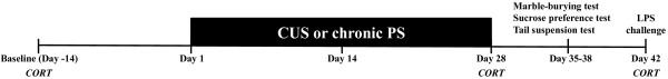
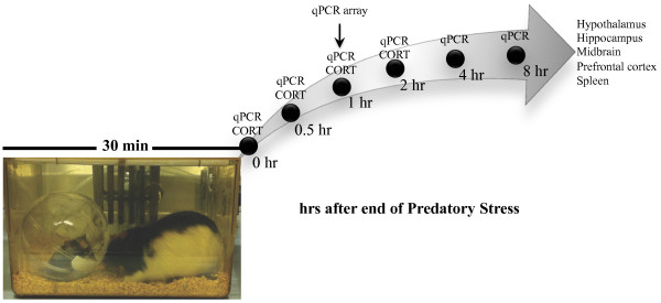
Methods: To address this, we have modified previously described models of rat/mouse predatory stress (PS) to increase the intensity of the interaction. We postulated that these modifications would enhance the predator-prey experience and increase neuroinflammation and behavioral dysfunction in prey animals. In addition, another group of mice were subjected to a modified version of chronic unpredictable stress (CUS), an often-used model of chronic stress that utilizes a combination of stressors that include physical, psychological, chemical, and other. The CUS model has been shown to exacerbate a number of inflammatory-related diseases via an unknown mechanism. Using these two models we sought to determine: 1) whether chronic PS or CUS modulated the inflammatory response as a proposed mechanism by which behavioral deficits might be mediated, and 2) whether chronic exposure to a pure psychological stressor (PS) leads to deficits similar to those produced by a CUS model containing psychological and physical stressors. Finally, to determine whether acute PS has neuroinflammatory consequences, adult mice were examined at various time-points after PS for changes in inflammation.

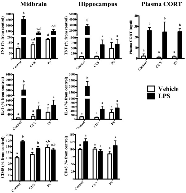
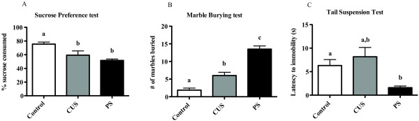
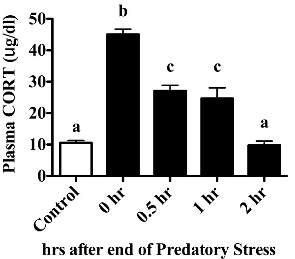
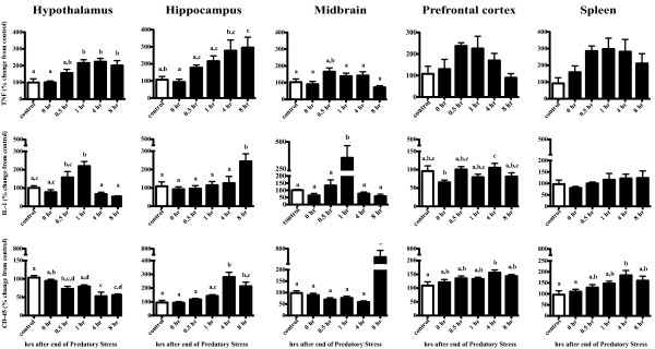
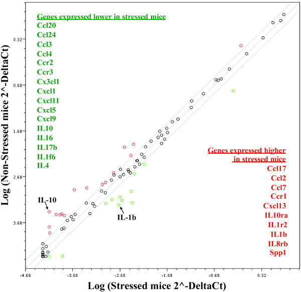
Results: Adolescent mice subjected to chronic PS had increased basal expression of inflammation within the midbrain. CUS and chronic PS mice also had an impaired inflammatory response to a subsequent lipopolysaccharide challenge and PS mice displayed increased anxiety- and depressive-like behaviors following chronic stress. Finally, adult mice subjected to acute predatory stress had increased gene expression of inflammatory factors.

Conclusion: Our results demonstrate that predatory stress, an ethologically relevant stressor, can elicit changes in neuroinflammation and behavior. The predatory stress model may be useful in elucidating mechanisms by which psychological stress modulates diseases with an inflammatory component.


Figures

Figure 1
Figure 1
Schematic and timeline for chronic stress. Mice were subjected to PS, CUS, or no stress (controls) for 28 consecutive days. To determine whether chronic stress resulted in habituation of the plasma CORT response, blood was collected from the facial vein 2 weeks prior to the first day of stress (Day -14), and 15 min after stress on Day 28. For non-stressed control mice, blood was taken on same days as above. The marble burying, sucrose preference, and tail suspension test were conducted 7-10 days following the final day of stress. To determine whether chronic stress modulated the inflammatory response to a subsequent challenge, mice were injected with 7.5 × 10⁵ EU/kg LPS at Day 42 and tissue was processed for analysis of gene expression.
Figure 2
Figure 2
Schematic and timeline for acute predatory stress model studies. PS consisted of placing a mouse inside a clear plastic hamster ball and then into the home cage of a large adult male Long Evans rat for 30 min. Mice were then examined for changes in plasma CORT and inflammatory mRNA within various brain regions (hypothalamus, hippocampus, midbrain, prefrontal cortex) and spleen immediately (0 min), 0.5, 1, 2 hr after PS ended. Inflammatory mRNA was also examined at 4 and 8 hr after PS. In a separate set of mice and to determine whether PS modulated a wider number of inflammatory genes in the midbrain, a qPCR array of 84 inflammatory genes was run at 1 hr post stress.
Figure 3
Figure 3
CUS and chronic PS lead to a suppressed inflammatory response to subsequent LPS challenge. Mice were subjected to 28 days of daily CUS or chronic PS and given an LPS challenge 14 days after the final day of stress. A 2 (LPS treatment) × 3 (Stress) ANOVA revealed that the inflammatory response to LPS was blunted (compared to controls) in mice subjected to CUS and chronic PS within the midbrain (TNF, IL-1) and hippocampus (TNF, CD45, IL-1). Within the midbrain of chronic PS mice, however, LPS did elicit a significant increase in TNF mRNA compared to chronic PS mice treated with saline (p < 0.05). A trend for an increase in IL-1 mRNA was also observed in CUS and chronic PS mice within the midbrain but did not reach statistical significance; p = 0.08 and p = 0.07, respectively. Additionally, while there was a tendency for chronic stress to increase basal levels of inflammation, this did not reach significance; midbrain (TNF & IL-1, p = 0.07) and hippocampus (IL-1, p = 0.08). Furthermore, while LPS increased plasma CORT levels in all LPS treated mice, no interaction with stress was observed. Data are expressed as percent change from control and presented as Mean ± SEM. Columns that do not share the same letter are significantly different (Two-way ANOVA p < 0.05). n = 6-8/group.
Figure 4
Figure 4
CUS and chronic PS increase depressive-, anhedonic-, and anxiety-like behaviors. Mice were subjected to 28 days of daily CUS or chronic PS and examined for changes in behavior 7-10 days later. A one-way ANOVA was used to examine potential differences between groups in all tests. (A) For anhedonia, mice were given a 48 hr two-bottle (water or 2% sucrose) choice to determine whether CUS or chronic PS altered sucrose preference. Control mice showed a significantly greater preference for sucrose compared to CUS and chronic PS mice. To examine whether anxiety was modulated by stress, we employed the marble-burying test. (B) Control mice showed reduced anxiety-like behavior and buried fewer marbles in a 30 min session compared to mice subjected to CUS and chronic PS mice. Additionally, chronic PS mice buried significantly more marbles than CUS mice. In a test of depressive-like behavior (tail-suspension test), chronic PS mice were faster to immobility than control and CUS mice (C). Data are expressed as Mean ± SEM. of grams of% sucrose consumed (A), number of marbles buried (B), or latency (seconds) to immobility. Columns that do not share the same letter are significantly different (One-way ANOVA; p < 0.05). Sucrose preference (n = 6-9/group), tail suspension (n = 10-16/group), marble burying (n = 9-16/group).
Figure 5
Figure 5
PS elicits an increase in plasma CORT that remains elevated for an hour following cessation. Mice were subjected to 30 min of PS and examined for changes in plasma CORT immediately (0 hr), 30 min (0.5 hr), 1 hr, or 2 hr after cessation of the stressor. A one-way ANOVA followed by Tukey's post hoc revealed that PS increased plasma CORT at 0, 0.5, and 1 hr compared to control mice and mice examined 2 hr after PS. Columns that do not share the same letter are significantly different (One-way ANOVA; p < 0.05). Data are expressed as Mean ± SEM. n = 8-12/group.
Figure 6
Figure 6
Gene expression analyses indicate increases in Th1 cytokines and microglia activation in multiple brain regions. To determine the extent that acute PS stimulated an inflammatory response, we conducted a time course examination of 4 brain regions and spleen. A one-way ANOVA and Tukey's post hoc revealed that both TNF and IL-1 were increased at respective time points following PS within the hypothalamus, hippocampus, and midbrain. Changes in the microglial activation marker CD45 were also increased within the hippocampus and midbrain, although not until 4 and 8 hrs after cessation, respectively. Changes in the prefrontal cortex and spleen are also provided. Columns that do not share the same letter are significantly different (One-way ANOVA; p < 0.05). Data are expressed as percent change from control and presented as Mean ± SEM. n = 6-8/group.
Figure 7
Figure 7
RT-PCR analysis of inflammation-related genes reveals qualitatively different response to PS in mouse midbrain. Mice were subjected to acute PS or remained in their home cage (control). 1 hr after acute PS, midbrain was dissected and examined for potential changes in inflammation. Samples were pooled together from 8 mice per genotype and run as n = 2 for both control and stressed mice. Mice subjected to stress show a > 2-fold increase in inflammatory factor gene expression presented in red text and > 2-fold decrease in inflammatory factors (green text) compared to control mice.

References

    1. You Z, Luo C, Zhang W, Chen Y, He J, Zhao Q, Zuo R, Wu Y. Pro- and anti-inflammatory cytokines expression in rat's brain and spleen exposed to chronic mild stress: involvement in depression. Behavioural brain research. 2011;225:135–141. doi: 10.1016/j.bbr.2011.07.006. - DOI - PubMed
    1. Bose M, Oliván B, Laferrère B. Stress and obesity: the role of the hypothalamic-pituitary-adrenal axis in metabolic disease. Current opinion in endocrinology, diabetes, and obesity. 2009;16:340–346. doi: 10.1097/MED.0b013e32832fa137. - DOI - PMC - PubMed
    1. Gu H, Tang C, Peng K, Sun H, Yang Y. Effects of chronic mild stress on the development of atherosclerosis and expression of toll-like receptor 4 signaling pathway in adolescent apolipoprotein E knockout mice. Journal of biomedicine & biotechnology. 2009;2009:613879. - PMC - PubMed
    1. Carroll J, Iba M, Bangasser D. Chronic Stress Exacerbates Tau Pathology, Neurodegeneration, and Cognitive Performance through a Corticotropin-Releasing Factor Receptor-Dependent Mechanism in a Transgenic Mouse Model of Tauopathy. The Journal of. 2011. - PMC - PubMed
    1. Danese A, Moffitt TE, Harrington H, Milne BJ, Polanczyk G, Pariante CM, Poulton R, Caspi A. Adverse childhood experiences and adult risk factors for age-related disease: depression, inflammation, and clustering of metabolic risk markers. Archives of pediatrics & adolescent medicine. 2009;163:1135–1143. doi: 10.1001/archpediatrics.2009.214. - DOI - PMC - PubMed

Publication types

MeSH terms